Glomus tumors are typically benign lesions that most often arise in the extremities. They are derived from the glomus body which is physiologically involved in thermoregulation. Usually these tumors are found in the subungual area of the fingers and toes; they are rarely found of the genitalia. In this study, we report the third known case of a glomus tumor of the scrotal skin. Our patient was a 53-year-old man of Middle Eastern descent who was diagnosed with a glomus tumor after a scrotal lesion was removed in our office under local anesthesia. Histopathological evaluation revealed a well circumscribed, 1 cm nodule that stained positive for alpha smooth muscle myosin and vimentin, and negative for S-100 and CD-34. Treatment of glomus tumors is primarily surgical and diagnosis is made on final pathology; low recurrence rates have been reported.
Glomus tumor; Scrotum, Urology; Glomus body; Histopathology
Glomus tumors are rarely found on the genitalia; consequently, when scrotal lesions are considered, these tumors are often low on the differential diagnosis. Despite their rarity, a glomus tumor should be considered when a painful scrotal mass exhibits pinpoint hypersensitivity and paroxysmal pain. Glomus tumors have characteristic staining patterns on histopathology and local excision of these tumors can be performed with low rates of recurrence.
Glomus tumors are rarely found on the genitalia; consequently, when scrotal lesions are considered these tumors are often low on the differential diagnosis. Despite their rarity, a glomus tumor should be considered when a painful scrotal mass exhibits pinpoint hypersensitivity and paroxysmal pain. Glomus tumors have characteristic staining patterns on histopathology and local excision of these tumors can be performed with low rates of recurrence.
Glomus tumors are rare, usually benign, lesions that arise most often in the extremities. They are derived from the glomus body and have a propensity to be found in the fingers and toes, and often in the subungual area [1]. They typically demonstrate pinprick sensitivity, cold hypersensitivity and paroxysmal pain [2]. Only two glomus tumors of the scrotum have been reported and we report a third case of this rare tumor.
A 53-year-old man of Middle Eastern descent was referred by the dermatology service for evaluation and consideration of excision of a painful scrotal mass that had been present for about 10 months. Other than hypertension, he had no significant medical history. Physical examination revealed a small, non-inflamed subcutaneous nodule of the scrotum located just to the right of median raphe. The initial impression was a sebaceous cyst; however, it was extremely tender to palpation. After his initial visit, the lesion was removed under local anesthesia in our office.
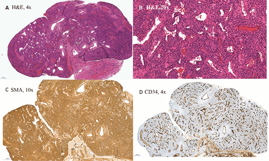
Grossly, the mass was a well circumscribed, 1 cm in size, and tan in color. The histopathological findings were consistent with a glomus tumor: positive for SMA and vimentin, and negative for S-100 and CD-34. (Figure 1)

Figure 1: (A) Sections shows a low power view with a distinct circumscription; (B) section showing sheets of Glomus (cuboidal) cells with a prominent “hemangiopericytomatous like” vascularity; (C) immunostains for SMA are positive; (D) CD34 highlighting a prominent “hemangiopericytomatous like” vascularity.
Glomus tumors are rare soft tissue neoplasms that are usually benign in nature [1]. They were first clinically described in 1877, and Masson described the microscopic appearance in 1924 [3]. The tumors are most commonly found in peripheral soft tissues such as the fingernail and toes, as these are the locations where glomus bodies are most prominent. In rare circumstances, these have been described to be found within the airway and stomach [1] and in the urogenital tract, the glans penis, urethra, bladder and kidney [4,5].
Glomus bodies consist of myoarterial shunts which are physiologically involved in thermoregulation. They consist of an arteriole anastomosed with a venule and they are located within the stratum reticularis of the dermis [6]. Diagnosis of a glomus tumor is primarily clinical; therefore, a thorough history and physical examination is paramount. Glomus tumors are most often bluish in hue, hypersensitive to cold temperatures, and exhibit local point tenderness. This tenderness can be demonstrated in the office using a Love’s test, which is performed by using a pinhead or pencil tip to elicit an intense pain [7]. Treatment for a glomus tumor is primarily surgical, as no effective medical therapy exists [8] and diagnosis is confirmed on histopathology. Since they are derived from modified smooth muscle, glomus tumors stain positive for SMA, and vimentin, but do not stain for S-100 and CD-34 [9]. Due to their relative rarity, the follow-up of patients with glomus tumors is not well described. Tumor recurrence, while unusual, has been reported [10].
The differential diagnosis of a glomus tumor includes other perivascular tumors including myopericytoma, myofibroma, and angioleiomyoma [11]. While these tumors may have overlap of their histopathological staining patterns, glomus tumors demonstrate a unique morphology of round cells exhibiting punched-out nuclei, pale cytoplasm, and a lacework of basement membrane material [6]. These pathological features are not demonstrated in the aforementioned tumors, and furthermore, glomus tumors are the only perivascular tumors found to be consistently painful in the clinical setting.
Of the now three reported cases of glomus tumor of the scrotum, each occurred in middle aged men [12,13]. This is the second case in a patient of Middle Eastern descent and, perhaps coincidentally, all three lesions were found to be in the right hemiscrotum. All tumors presented in relatively small size, being 1 cm or less. Our case included, no recurrence of these tumors has been reported.
Glomus tumors of the scrotum are rare entities; however, the diagnosis should be considered when a non-inflammatory superficial genital mass exhibits point tenderness and cold hypersensitivity. Treatment involves surgical excision, and diagnosis is confirmed on final histopathology.
- Mravic M, LaChaud G, Nguyen A, Scott MA, Dry SM, et al. (2015) Clinical and histopathological diagnosis of glomus tumor: an institutional experience of 138 cases. Int J Surg Pathol. 23:181-188. [Crossref]
- Kim SW, Jung SN. (2011) Glomus tumour within digital nerve: a case report. J Plast Reconstr Aesthet Surg. 64:958-960. [Crossref]
- Masson P (1924) Le glomus neuromyoarterial des regions tactiles et ses tumeurs. Lyon Chir. 21:257-280. [Crossref]
- Dagur G, Warren K, Miao Y, Singh N, Suh Y, et al. (2016) Unusual glomus tumor of the penis. Curr Urol. 9:113-118. [Crossref]
- He T, Hu J, Jin L, Li Y, Liu J, et al (2016). Glomus tumor of the anterior urethra: A rare case report and review of the literature. Mol clinical oncol. 4:1057-1059. [Crossref]
- Goldblum J, Enzinger FM, Weiss SW, Folpe AL (2014). Enzinger and Weiss’s soft tissue tumors. (pp. 749-765). Philadelphia, PA: Saunders/Elsevier. [Crossref]
- Love JG (1944) Glomus tumors: diagnosis and treatment. Proc Staff Meet, Mayo Clin. 19:113-116. [Crossref]
- Hazani R, Houle J, Kasdan ML, Wilhelmi BJ (2008) Glomus tumors of the hand. Eplasty. 8:e48. [Crossref]
- Wang ZB, Yuan J, Shi HY. (2014) Features of gastric glomus tumor: a clinicopathologic, immunohistochemical and molecular retrospective study. Int J Clin Exp Pathol. 7:1438-1448. [Crossref]
- Grover C, Khurana A, Jain R, Rathi V. (2013) Transungual surgical excision of subungual glomus tumour. J Cutan Aesthet Surg. 6:196-203 [Crossref]
- Zhao M, Williamson SR, Sun K, Zhu Y, Li C, et al. Benign perivascular myoid cell tumor (myopericytoma) of the urinary tract: a report of 2 cases with an emphasis on differential diagnosis. Human Pathology. 45:1115-1121. [Crossref]
- Miyoshi H, Yamada N, Uchinuma E (2009) A glomus tumour of the scrotal skin. Plast Reconstr Aesthet Surg. 62:414-415. [Crossref]
- Khalafalla K, Al-ansari A, Omran A, Farghaly H, Alobaidy A (2017) Glomus tumor of the scrotum: a case report and mini-review. Curr Urol. 10:213-216. [Crossref]

