Long-standing mitral valve disease is associated with enlargement of the left atrium as a compensatory mechanism. Giant left atrium is seen in only 19% of patients and such a low incidence is probably related to an early development of pulmonary hypertension.
We report the case of a giant left atrium with big thrombus diagnosed during an echocardiography done systematically as part of an etiologic assessment of a stroke.
Mitral Valve; Stenosis; Thrombosis
Echocardiography is the procedure of choice for the diagnosis of cardiac mass involving the left atrium. It has been shown to be a superior method in defining the characteristics of a mass in the left atrium: as it helps to characterize the mass by morphologic shape and appearance, site of attachment, types of margins, and presence or absence in the left atrial appendage.
This is a 70-year-old woman without cardiovascular risk factors, having led four pregnancies without incidents, who has presented one month ago a regressive cerebral vascular accident. She was put by her neurologist under aspirin then addressed to the cardiology department for etiological assessment of her stroke. The patient complained no dyspnea or palpitation. Physical examination did not reveal signs of heart failure. At heart auscultation she had an apical diastolic rumble. Electrocardiography showed only an atrial fibrillation. A hugely enlarged left atrium (80 cm2 / 140 ml/m2) occupied by a big thrombus (70 × 60mm) was demonstrated by transthoracic echocardiography (Fig. 1).
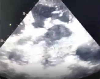
Figure 1. 2D-TTE: four chamber incidence huge left atrial thrombus upstream a mitral stenosis
Evidence of calcified rheumatic mitral stenosis (mitral valve area was about 0.7 cm2: measured with planimetry technique) (Fig. 2) with mild regurgitation was seen.
The two mitral commissures were fused and the sub-mitral valve was retracted.
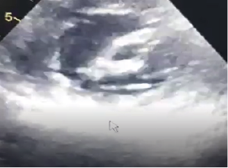
Figure 2. 2D-TTE short axis: mitral orifice stenosis
Tricuspid valve was severely incompetent with an estimated pulmonary artery systolic pressure of 80 mmHg. The tricuspid ring was dilated to 24 mm / m2. The aortic valve was also affected by rheumatism, it was remodeled and the site of minimal aortic insufficiency (Fig. 3).
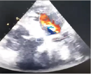
Figure 3. 2D: TTE 4 chamber view: mild aortic regurgitation
After performing coronarography which was normal the patient was addressed in emergency to heart surgery department for mitral valvular replacement with tricuspid annuloplasty associated with surgical thrombectomy.
The terminology of huge left atrium is for diameters larger than 6 cm, and a left atrium with a diameter of more than 12 cm is very rarely reported in the literature [1]. The causes of left atrium enlargement include mitral stenosis or regurgitation, rheumatic valve, left ventricular failure, hypertension, left ventricular diastolic dysfunction and atrial fibrillation [2]. Long standing mitral valve disease is associated with enlargement of the left atrium as a compensatory mechanism due to increase intracavitary pressure and volume. Such an enlargement is beneficial as it reduces pulmonary congestion, thus it protects the lung from pulmonary hypertension and oedema [3]. The aetiology of huge left atrium (LA) is still unknown. The mechanism of formation of giant left atrium (GLA) is not fully understood. Patients with chronic mitral valve disease are not always associated with GLA. Only 19 % may develop such a condition [4]. Previous studies showed that chronic pressure in the left atrium is not the only cause of GLA, but weakening of the left atrial wall by rheumatic pancarditis causing chronic inflammation and fibrosis is also implicated [5]. Enlargement of the left atrium is associated with development of atrial fibrillation, which in return can lead to further enlargement of left atrium. Huge enlargement of the left atrium is prone to develop various complications including thrombus formation, thromboembolic events [6]. The enlarged left atrium is associated with blood stasis and thrombus formation. The risk of thromboembolism increases with left atrial size regardless of anticoagulation [7]. LA dilation leads to thinning and fibrosis of myocardial and conducting fibers and thereby results in electrical tissue inhomogeneity, disordered electrical activation and contractility. Every 5 mm increase in LA diameter increases the development of atrial fibrillation by 39% [8-10].
Previous studies found that the incidence of left atrial thrombus among patients with mitral stenosis associated with AF varies from 7-38%. Such an incidence is directly related to the size of left atrium [11]. It is interesting to note that not all patients with GLA have an associated thrombus formation [12]. Goldsmith and colleagues , found that patients with mitral valve disease have associated left atrial endocardial damage [13]. Such damage contributed to the risk of thrombus formation. The other factor is related to the activation of coagulation system within the left atrium [14]. Patients with mitral stenosis have a significantly high level of fibrinopeptide A, thrombin-antithrombin III complex and Von Willebrand factor antigen in the left atrium. These biochemical markers are responsible for the formation of thrombosis even during anticoagulation regardless of the severity of mitral stenosis or size of left atrium. Our patient had a mitral stenosis associated with chronic AF. Both factors were responsible for the development of Giant Left Atrium and thrombosis. In such situation medical treatment is based on effective anticoagulation. The aim of surgery in giant left atrium is to correct the mitral valve abnormalities, to treat compressive manifestations, to prevent thromboembolism and to revert atrial fibrillation into normal sinus rhythm especially by associating plicature of left atrium at the mitral valve replacement. Plication of the left atrium was not indicated in our patient, to avoid unnecessary complication as circumflex coronary artery injury, pulmonary vein obstruction and oesophageal stricture. Previous study by Tonguc and colleagues [15], compared patients undergoing mitral valve replacement (MVR) with or without plication, found no significant difference in hemodynamic improvement and reduction of left atrial diameter .Also, the incidence of left atrial thrombus after MVR showed no difference whether plication was performed or not [15]. If the thrombus is big and organized thrombectomy is indicated however removal of an organized thrombus from the left atrium can be challenging especially when it is huge in size.
Cardiac thrombi appear more frequently than cardiac tumours. They are typically located in the atrium, more often in the left, and generally occur in patients with organic heart disease especially mitral stenosis. Their treatment is often based on effective anticoagulation with K antivitamins. Surgical thrombectomy is rarely indicated, especially in the presence of large and organized ones.
- Kawazoe K., Beppu S., Takahara Y., Nakajima N., Tanaka K., et al. (1983) Surgical treatment of giant left atrium combined with mitral valvular disease. Plication procedure for reduction of compression to the left ventricle, bronchus, and pulmonary parenchyma. J Thorac Cardiovasc Surg 85: 885‑892. [Crossref]
- Cuspidi C., Negri F., Sala C., Valerio C., Mancia G (2012) Association of left atrial enlargement with left ventricular hypertrophy and diastolic dysfunction: a tissue Doppler study in echocardiographic practice. Blood Press 21: 24‑30. [Crossref]
- Schwammenthal E., Vered Z., Agranat O., Kaplinsky E., Rabinowitz B., et al. (2000) Impact of atrioventricular compliance on pulmonary artery pressure in mitral stenosis: an exercise echocardiographic study. Circulation 102: 2378‑2384. [Crossref]
- Di Eusanio G., Gregorini R., Mazzola A., Clementi G., Procaccini B., et al. (1988) Giant left atrium and mitral valve replacement: risk factor analysis. Eur J Cardio-Thorac Surg Off J Eur Assoc Cardio-Thorac Surg 2: 151‑159. [Crossref]
- Hurst JW (2001) Memories of patients with a giant left atrium. Circulation 104: 2630‑2631. [Crossref]
- Goldstein LB (2011) Left atrial enlargement: A cause of stroke? CMAJ Can Med Assoc J 183: 1129‑1130. [Crossref]
- Apostolakis E., Shuhaiber JH (2008) The surgical management of giant left atrium. Eur J Cardio-Thorac Surg Off J Eur Assoc Cardio-Thorac Surg 33: 182‑190. [Crossref]
- Davies MJ., Pomerance A (1972) Pathology of atrial fibrillation in man. Br Heart J 34: 520‑525. [Crossref]
- Bailey GW., Braniff BA., Hancock EW., Cohn KE (1968) Relation of left atrial pathology to atrial fibrillation in mitral valvular disease. Ann Intern Med 69: 13‑20. [Crossref]
- Vaziri SM., Larson MG., Benjamin EJ., Levy D (1994) Echocardiographic predictors of nonrheumatic atrial fibrillation. The Framingham Heart Study. Circulation 89: 724‑730. [Crossref]
- Farman MT., Sial JA., Khan N., Rahu QA., Tasneem H., (2010) Severe mitral stenosis with atrial fibrillation--a harbinger of thromboembolism. JPMA J Pak Med Assoc 60: 439‑443. [Crossref]
- Darwazah AK., El Sayed H (2013) Giant left atrium associated with massive thrombus formation. Thromb J 11: 5. [Crossref]
- Goldsmith I., Kumar P., Carter P., Blann AD., Patel RL., et al. (2000) Atrial endocardial changes in mitral valve disease: a scanning electron microscopy study. Am Heart J 140: 777‑784. [Crossref]
- Yamamoto K., Ikeda U., Seino Y., Mito H., Fujikawa H., et al. (1995) Coagulation activity is increased in the left atrium of patients with mitral stenosis. J Am Coll Cardiol 25: 107‑112. [Crossref]
- Tonguç E., Kestelli M., Özsöyler İ., Yilik L., Yilmaz A., et al. (2001) Limit of Indication for Plication of Giant Left Atrium. Asian Cardiovasc Thorac Ann 9: 24‑26. [Crossref]