The arcuate uterus though a classified mullerian duct anomaly but is mild anatomic variant without any association with reproductive failure. The adenomyosis is abnormal ectopic location of endometrium in the myometrium which can be focal and diffuse, and assumes cystic appearance on repeated haemorrhages. The medical resonance imaging is important imaging modality to establish both the diagnosis and both the condition in the same patient is rare and not reported in the literature so far.
Arcuate uterus with cystic adenomyosis is a rare finding to be seen together and not reported in literature so far. The arcuate uterus is mild form of anatomic variation of uterus without any consequences on infertility outcome. Cystic adenomyosis due to repeated cyclic bleeding in the ectopic endometrial tissue in the myometrium. The MR imaging is the modality of choice for diagnosis of the both
We present a case of 20 year female presenting with Dysmenorrhoea MRI images showed normal external contour of uterus with broad based indentation of myometrium into the endometrium suggestive of arcuate uterus and there is also presence of focal cystic adenomyosis seen as focal cystic changes seen with variable signal on T1w and T2w images due to haemorrhage in different stages. Diagnosis is Arcuate Uterus with focal cystic adenomyosis.
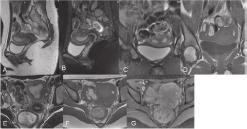
Figure 1. A 20 year female presenting with Dysmenorrhoea, MRI images (A-Sag T2W, B-Sag T2 FATSAT, C, D- Cor T2W and T1W E, F, G-axial T2w, T1W and Post contrast) showing normal external contour of uterus with broad based indentation of myometrium into the endometrium suggestive of arcuate uterus and there is also presence of focal cystic adenomyosis seen as focal cystic changes seen with variable signal on T1w and T2w images due to haemorrhage in different stages. Diagnosis is Arcuate Uterus with focal cystic adenomyosis
Arcuate uterus is myometrial fundal indentation into the endometrium which is >1cm and is most common anomaly of mullerian duct due to incomplete resorption of uterovaginal septum. Focal cystic adenomyosis is rare variation of adenomyosis which may have cystic spaces due to repeated haemorrhages. Arcuate uterus is least common Mullerian duct anomaly associated with infertility. Focal adenomyosis results in pelvic pain and dysmenorrhoea. Though the clinical consequences of both the entities i.e. arcuate uterus and focal cystic adenomyosis are of not much importance but their presence in single patient make it a rare entity and of good academic importance. Key imaging diagnostic clues
1. Arcuate uterus- Normal external contour of uterus with broad based indentation of myometrium into the endometrium
2. Focal Cystic Adenomyosis-Focal cystic changes seen with variable signal on T1w and T2w images due to haemorrhage in different stages.
- Mueller GC, Hussain HK, Smith YR, et al. (2007) Mullerian duct anomalies: comparison of MRI diagnosis and clinical diagnosis. AJR Am J Roentgenol 189: 1294-1302. [Crossref]
- Manta L, Suciu N, Constantin A, et al. (2016) Focal adenomyosis (intramural endometriotic cyst) in a very young patient-differential diagnosis with uterine fibromatosis. J Med Life 9: 180. [Crossref]
- Mazouni C, Girard G, Deter R, et al. (2008) Diagnosis of Mulleriananomalies in adults: evaluation of practice. Fertil Steril 89: 219-222. [Crossref]
- Sutton D (2003) Text Book of Radiology and Imaging. Churchill Livingstone. London 2: 1453-1487.
- Adam A, Dixon AK, Gillard JH, et al. (2014) Grainger & Allison's Diagnostic Radiology E-Book. Elsevier Health Sciences.
- Gupta AK, Garg A, Khandelwal N (2017) Diagnostic Radiology: Gastrointestinal and Hepatobiliary Imaging. JP Medical Ltd.
- Haaga JR, Boll D (2016) Computed Tomography & Magnetic Resonance Imaging of the Whole Body E-Book. Elsevier Health Sciences.
Editorial Information
Article Type
Case Report
Publication history
Received date: June 9, 2020
Accepted date: June 21, 2020
Published date: June 29, 2020
Copyright
©2020 Rana L. This is an open-access article distributed under the terms of the Creative Commons Attribution License, which permits unrestricted use, distribution, and reproduction in any medium, provided the original author and source are credited.
Citation
Rana L, Sood D, Rana N, Singh D (2020) Arcuate Uterus with Focal Cystic Adenomyosis-A Case Report. OSP J Case Rep 2. JCR-2-125
Corresponding author
Lokesh Rana
Assistant Professor, Department of Radio-diagnosis, Dr RPGMC, Kangra at Tanda. Himachal Pradesh, India. poojalokesh2007@gmail.com
Figure 1. A 20 year female presenting with Dysmenorrhoea, MRI images (A-Sag T2W, B-Sag T2 FATSAT, C, D- Cor T2W and T1W E, F, G-axial T2w, T1W and Post contrast) showing normal external contour of uterus with broad based indentation of myometrium into the endometrium suggestive of arcuate uterus and there is also presence of focal cystic adenomyosis seen as focal cystic changes seen with variable signal on T1w and T2w images due to haemorrhage in different stages. Diagnosis is Arcuate Uterus with focal cystic adenomyosis