Birds are considered a good model for studying the phonation process, the syrinx is a vocal organ in birds. The purpose of this study is to investigate the topographical and morphological characteristics of syrinx of male domestic fowl. In the current study we use the syrinxes of seven adult males. The study shows that the syrinx of investigated birds is tracheobronchial in type. It consists of; tympanum, tracheosyringeal and bronchosyringeal groups. In addition, there are interbronchial ligament (brachidesm), lateral and medial vibrating membranes as well as the pessulus at the tracheal bifurcation. Tympanum part forms the first part of the syrinx; it is formed of four tracheal rings. The tracheosyringeal part is located at the point of tracheal bifurcation just below the tympanum. It is formed of four highly modified incomplete tracheal rings. The bronchosyringeal part is formed of first three pairs of bronchial half-rings. The current study was presented the detailed morphological characteristics of syrinx in male domestic fowl.
The syrinx is the organ of voice in birds and is probably present in all avian species. Some birds vocalize all year long while others call only during the mating season or during migration [1]. According to Baumel et al. [2], there are three types of syrinx; tracheal, tracheobronchial and bronchial, depending on the deviation of the cartilages of the syrinx from either the trachea or the bronchi. The tracheal elements laid cranial to the bifurcation of the airway, while the bronchial elements laid at the most cranial part of the right and left primary bronchi. The great majority of birds are described as having a tracheobronchial syrinx which is located at the bifurcation of the trachea as stated by Nickel et al. [3], the tracheobronchial syrinx has both tracheal and bronchial elements. The classification of birds depended on presence or absence of the basic structure of syrinx or their musculature [4]. The regulation of airflow and action influences acoustic parameters of syrinx is controlled by muscles during closing and opening of the passage airways [5-7].
There is lack of information concerning the anatomical features of the syrinx in male domestic fowl. So the aim of the present study is to give more information about the morphological features of syrinx of male domestic fowl. But why is this important?
Seven males of domestic fowl collected from Assiut governorate (weighted about 1450 to 1500 gm) were used in this study. All of the birds were slaughtered. After thoracic incision, the topographical position of syrinx was observed, and the anatomical examination was done. Different measurements were taken using Precision Digital Vernier Caliper (the length of tracheolateralis and sternotrachealis muscles). The syringes of cocks were left in 1% methylene blue solution for 15 min then passed through 70% alcohol for two hours for the cartilages to become more evident and then photographed. The syringes were observed, described and photographed using a stereomicroscope (LEICA S6D) fitted with a digital camera (DFC-290).
The syrinx of examined cocks is observed within the body cavity. It lies ventral to the esophagus and dorsal to the base of the heart. It is a tracheobronchial in type and consists of three groups of rings: cranial (tympanum) group, intermediate (tracheosyringeal) group and caudal (bronchosyringeal) group. In addition, there are interbronchial ligament (brachidesm), lateral and medial vibrating membranes as well as the pessulus at the tracheal bifurcation.
The tympanum part forms the first part of the syrinx. It is formed of four tracheal rings, firmly attached together by dense fibrous tissue, especially along their sides. They are difference in size and shape between the tracheal rings immediately above it and intermediate syringeal cartilages immediately below it (Figures 1-4).

Figure 1. In situ ventral view of the syrinx in the domestic male fowl. 1. Trachea; 2. Tympanum; 3. Tracheolateralis muscles; 4. Sternotrachealis muscles; 5. Sternum; 6. Heart
The tracheosyringeal part is located at the point of the tracheal bifurcation just below the tympanum. It is formed of four highly modified incomplete tracheal rings, compressed laterally. They are fused with the pessulus ventrally, and their free ends directed dorsally. The first intermediate syringeal ring is smaller than the other three (Figure 3&4A).
The bronchosyringeal part is formed of the first three pairs of bronchial half-rings, on each side of the syrinx and their free ends directed dorsomedially. The first bronchosyringeal half-rings are modified and larger than the other three and attached at the both ends to the pessulus. The second bronchosyringeal half-rings are only slightly modified, their ventral ends attached ventrally to the first bronchosyringeal half-rings while their dorsal ends were free. The third bronchosyringeal half-rings are much similar to the bronchial half-rings which lie immediately caudal to them (Figures 2-4).
The pessulus is triangular in shape, located at the summit of the bronchial junction. Its pointed apex directs cranially through the lumen of syrinx, while its broad base directs caudally (Figures 2-4).
Two pairs of lateral and medial tympaniform membranes are observed. The lateral tympaniform membranes (Figures 2-4A) are curved inward through the syringeal lumen, from the last intermediate syringeal to the first bronchosyringeal half-rings. The medial tympaniform membranes are stretched between the free ends of bronchosyringeal half-rings and extended caudally from the base of the pessulus to the level of the third bronchosyringeal half-rings (Figures 2&3).
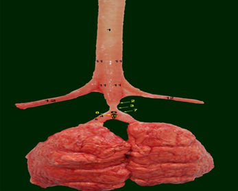
Figure 2. Dorsal view of the syrinx in the domestic male fowl, in fresh state. 1. Trachea; 2. Tympanum; 3. Tracheosyringeal rings 4. Bronchosyringeal half-rings; 5. Bronchus; 6. Pessulus; 7. Lateral tympaniform membrane 8. Medial tympaniform membrane; 9. Interbronchial ligament; 10. Interbronchial foramen; 11. Tracheolateralis muscle; 12. Sternotrachealis muscle

Figure 3. lateral view of the syrinx in the domestic male fowl, in fresh state. 1. Trachea; 2. Tympanum; 3. Tracheosyringeal rings 4. Bronchosyringeal half-rings; 5. Bronchus; 6. Pessulus; 7. Lateral tympaniform membrane 8. Medial tympaniform membrane; 9. Interbronchial ligament; 10. Interbronchial foramen; 11. Tracheolateralis muscle; 12. Sternotrachealis muscle

Figure 4. Gross anatomic appearance of the syrinx of male domestic fowl; lateral (A), dorsal (B) and ventral (C) views. (A-C): Tympanum rings (black stars), Tracheosyringeal rings (arrows), Bronchosyringeal half-rings (white stars), Lateral tympaniform membrane (arrowheads)
The intrinsic syringeal muscles are absent, but the extrinsic syringeal muscles are seen. The paired tracheolateralis (TL) muscles are detected laterally at the both sides of the trachea and extended from the larynx to the cranial part of the tympanum (Figures 2&3). The paired sternotrachealis (ST) muscles are extended from the medial aspect of the xiphisternal processes of the sternum to the lateral aspects of the caudal part of the trachea (Figures 1-3). ST muscles are fused laterally with TL muscles and both covered the trachea dorsally and laterally. The left ST and TL muscles are well developed than the right muscles. The length of the TL muscle ranges between 10.5- 15 cm, while ST muscle ranges between 1.5-3 cm.
The interbronchial ligament (Figures 2&3) connecting the right and left primary bronchi at the end of the medial tympaniform membranes. It extends from the third bronchosyringeal to fifth bronchial half-rings. The interbronchial foramen (Figures 2&3) is observed between pessulus, interbranchial ligament and medial tympaniform membranes.
The topographical findings of the syrinx in male adult fowl in this study were similar to those of mallards [8,9] geese [10,11] Denizli roosters [12] pigeons [13,14] turkeys [15-17] guinea fowls [18] and ostriches [19]. It could be classified as tracheobronchial in type as described in most common birds such as pigeons [14] quails [20,21] mallards [8,9,22] geese [10,11] ostriches [19] long legged buzzards [23] Denizli roosters [12] guinea fowls [18] black francolins [24] and eagle owls [21].
The present results revealed that the tympanum was consisted of four tracheal rings, like the Denizli roosters [12] and sparrow hawks [25]. The finding obtained from this investigation was in agreement with Myers and Meclelland [26,27] and dissimilar from the result of Freeman Dyce et al., and King [28-30] which recorded that the tympanum was consisted of three or five tracheal rings in chickens. While the number of tracheal rings forming the tympanum were reported as two in quails [20,21], three in long legged buzzards [23] ostriches [19] guinea fowls [18] turkeys [31] and song birds [32] five in sea gulls [33] and pigeons [14].
The present investigation recorded four highly modified tracheosyringeal (intermediate) incomplete rings, opened dorsally and attached to the pessulus ventrally, which was in agreement with Myers and Meclelland [26,27]. As reported in pigeons and song birds, there were four C-shaped intermediate syringeal rings [14,27,34,35] while in the ostriches only last two rings were C-shaped and the first two were circled [46]. Two C-shaped intermediate rings were reported in guinea fowls [18] black francolins [24] and turkeys [31] two-three C-shaped intermediate rings in long legged buzzards [23].
The bronchosyringeal part was consisted of first three pairs of bronchial half-rings in this investigation, which was in agreement with Myers, Meclelland, King and Mclelland [26,27,34]. Similar observation was also recorded in song birds [19] ostriches [32] guinea fowls [18] Denizli rosters [12] and turkeys [31]. It has been reported that it was consisted of first two bronchial half-rings as in quails [20,21] first four bronchial half-rings as in black francolins [24] and long-legged buzzards [23] first five half-rings as in pigeons [14] first five-six half-rings as in herring gulls [35] first six half-rings as in geese [9-11] first seven half-rings as in sea gulls [33].
In the present study, the pessulus was triangular in shape and its pointed apex directed cranially, which was in agreement with Myers and Meclelland [26,27]. Similar results were also revealed in sea gulls [33] black francolins [24] songbirds [21,32] long legged buzzards [23] turkeys [37,39] and guinea fowls [2,18]. On the other hand, it was absent as in penguins [12,20] larks [35,38] and pelicans [39].
The lateral and medial tympaniform membranes located at syrinx undertake the voice production, these results entirely confirmed with authors in several types of birds [2,35,40-42]. The location of lateral tympani form membrane in this investigation was the same with Myers [26] Bell and Freeman [43] as well as Meclelland [27] similar location also reported in ostriches [19] singing birds [32] and turkeys [15,17]. While different locations were reported in some birds such as; it was extended between the third and fourth tracheosyringeal rings as in pigeons [14,27,44] located between first and second bronchosyringeal half-rings as in sea gulls [2,33] and long legged buzzards [23]. The inward curve of that membrane which gave the syrinx its characteristic appearance was also recorded by King and Mclelland and Nickel et al. [3,34]. The location of medial tympaniform membrane in the present work was in agreement with Myers and Meclelland [26,27] similar observation was also recorded in ostriches [19] Japanese quails [21] Denizli roosters [12] mallards [9,22] and song birds [43]. This observation different from the result of Onuk et al. [10], in geese, which recorded that it was connected the pessulus and second bronchosyringeal half-ring.
The present study revealed that the extrinsic muscles were sternotrachealis and tracheolateralis muscles, which was in agreement with Brackenbury and Meclelland [27,45]. Our observations were partly different from those of Myers and Meclelland [26,27]. Myers [26] revealed that the ST muscles were the only extrinsic muscles in domestic fowl, it extended from the anteriolateral process of the sternum to the trachea and both muscles were proceed along the sides of the trachea to their final insertion on the ventral cartilages of the upper larynx. While Meclelland [27] stated that the ST muscles were combined with TL muscles to form a composite muscle which enclosed the trachea except dorsally. The ST and TL muscles were also recorded in many birds such as turkeys [17] pigeons [14,27] francolins [24] ducks [22] sea gulls [46] eagle owls [21] and parrots [43] while Yildiz et al. [19], reported that the TL muscles were absent in ostriches. The intrinsic muscles were not observed in this investigation, while those muscles were recorded in songbirds and parrots [43].
There was interbronchial ligament reported in the present investigation, which observed also in ostriches [19] turkeys [17,37] geese [10] guinea fowls [18] long legged buzzards [23] sea gulls [33] black francolins [24] many singing birds [3,12,32] and mallards [9,28,27]. On the other hand, it was absent as reported in pigeons [14].
- Zhang K, Siegmund T, Chan RW, Fu M (2009) Predictions of fundamental frequency changes during phonation based on a biomechanical model of the vocal fold lamina propria. J Voice. 23: 277-282. [Crossref]
- Baumel JJ, King AS, Breazile JE, Evans HE, Berge JC (1993) Handbook of Avian Anatomy: Nomina Anatomica Avium. 2nd edn. [Crossref]
- Nickel R, Schummer A, Seiferle E (1977) Anatomy of the Domestic Birds. 2nd edn.
- Berlin. Hamburg. Parey; Germany. [Crossref]
- Tsukahara N, Yang Q, Sugita S (2008) Structure of the syringeal muscles in jungle crow (Corvus macrorhynchos). Anat Sci Int. 83: 152-158. [Crossref]
- Andersson M (1994) Sexual Selection Princeton. NJ: Princeton University Press. [Crossref]
- Podos J, Huber SK, Taft B (2004) Bird song: the interface of evolution and mechanism. Annu Rev Ecol Evol Syst. 35: 55-87. [Crossref]
- Edwards SV, Kingan SB, Calkins JD, Balakrishnan CN, Jennings WB, et al (2005) Speciation in birds: genes, geography, and sexual selection. PNAS. 102: 6550-6557. [Crossref]
- Frank T, Probst A, Konig HE, Walter I (2007) The syrinx of the male mallard (Anas platyrhynchos): special anatomical features. Anat Histol Embryol. 36: 121-126. [Crossref]
- Yilmaz B, Yilmaz R, Arican I, Yildiz H (2012) Anatomical Structure of the Syrinx in the Mallard (Anas platyrhynchos). Harran Univ Vet Fak Derg. 1: 111-116. [Crossref]
- Onuk B, Haziroglu RM, Kabak M (2010) The Gross Anatomy of Larynx, Trachae and Syrinx in Goose (Anser anser domesticus). Kafkas Univ Vet Fak Derg. 16: 443-450. [Crossref]
- Mohamed R (2017) Gross anatomical syringeal structures of goose (Anser anser domesticus). J Adv Vet Anim Res. 4: 343-347. [Crossref]
- Taşbaş M, Hazıroğlu RM, Çakır A, Özer M (1994) Morphological investigations of the respiratory system of the Denizli cock. II. Laryx, trachea, syrinx. Ankara Univ Vet Fak Derg. 41: 135-153.
- Warner RW (1972) The syrinx in family Columbidea. J Zool. 166: 385-390. [Crossref]
- Yildiz H, Yilmaz B, Arican I (2005) Morphological Structure of the Syrinx in the Bursa Roller Pigeon (Columba livia). Bull Vet Inst Pulawy. 49: 323-327. [Crossref]
- Cover MS (1953) Cross and microscopic anatomy of the respiratory system of the turkey. Am J Vet Res. 230-238. [Crossref]
- Arican I, Yildiz H, Yilmaz B (2007) Morphometric Studies on Vocal Organ of White Turkey. Indian Vet J. 84: 964-966.
- Khaksar Z, Kookhdan ET, Parto (2012) A study on anatomy and histological structure of Larynx in male and female turkeys. World J Zool. 7: 245-250. [Crossref]
- Al-Bishtue AH. (2014) Anatomical investigations of the syrinx (voice box) of the adult male West African guinea fowl (Numida meleagris galeata) in the Al-Najaf Al- Ashraf province Al-Qadisiya . J Vet Med Sci. 13: 100-105. [Crossref]
- Yildiz H, Bahadir A, Akkoc A (2003) A study on the morphological structure of syrinx in ostriches (Struthio camelus). Anat Histol Embryol. 32: 187-191. [Crossref]
- Bayram G, Liman N (2000) A morphological investigation of the postnatal development of the syrinx in the quail. Turk J Vet Anim Sci. 24: 381-392. [Crossref]
- Cevik-Demirkan A, Haziroglu RM, Kurtul I (2007) Gross morphological and histological features of larynx, trachea and syrinx in Japanese quail. Anat Histol Embryol. 36: 215-219. [Crossref]
- Mohamed R (2017) Sexual Dimorphism in the Anatomical Features of the Syrinx in the White Pekin Ducks (Anas platyrhynchos). 5: 78-85. [Crossref]
- Kabak M, Orhan IO, Haziroglu RM (2007) The gross anatomy of larynx, trachea and syrinx in the long-legged buzzard (Buteo rufinus). Anat Histol Embryol. 36: 27-32. [Crossref]
- Al-aameli M, Kadhim kK. (2017) Histomorphological Study of Syrinx of Black Francolin (Francolinus francolinus) in Iraq. Adv Anim Vet Sci. 5: 92-99. [Crossref]
- Ozudogru Z, Balkaya H, Kara A, Ozdemir D (2015) A Study of the Morphological Structure of the Syrinx of the Sparrowhawk (Accipiter nisus). Isr J Vet Med. 70. [Crossref]
- Myers JA (1917) Studies on the syrinx of Gallus domesticus. J Morphol. 20: 165-215. [Crossref]
- Meclelland J (1990) A colour Atlas of Avian Anatomy. Wolfe publishing Ltd; England. [Crossref]
- Freeman B (1983). Physiology and Biochemistry of the Domestic Fowl. Academic Press. 234-256.
- Dyce KM, Sack WO, Wensing CJ (1987) Textbook of Veterinary Anatomy. 5th edn. London. UK: Saunders. [Crossref]
- King AS (1989) Functional anatomy of the syrinx. In: Form and Function in Birds. Academic Press: London.
- Ragab AS, Reem RT, Rezk MH, Nora AS (2016) The Gross Anatomy of the syrinx of the turkey. Int J Adv Res Biol Sci. 3: 82-90. [Crossref]
- Warner RW (1972). The anatomy of the syrinx in passerine birds. J Zool Lond. 168: 381-393. [Crossref]
- Ince NG, Pazvant G, Alpak H (2012) Anatomical features of the syrinx in sea gulls. Ankara Üniv Vet Fak Derg 59: 1-3. [Crossref]
- King AS, Mclelland J (1975) Outlines of Avian Anatomy. 1st edn. Bailiere and Tindall; London. [Crossref]
- King AS, Mclelland J (1984) Birds Their Structure and Function. 2nd edn: Bailiere Tindall; London. [Crossref]
- Hartley RS, Suthers RA (1990) Lateralvation of synngealfunction during song production in the canary. L Neurobiol. 2: 1236-1248.
- AL-Mussawy AM (2011) Anatomical and Histological Study of Major Respiratory Organs (Larynx, Trachea, Syrinx, Bronchi and Lungs) In Indigenous Male Turkey (Meleagris gallopava). AL-Qadisiya. Uni Vet Med College. [Crossref]
- Ames LP (1971) The Morphology of the Syrinx in Passerine Birds. Bulletin Peabody Museum of Natural History. [Crossref]
- Griffiths CS (1994) Monophyly of the Falconiformes based on syringeal morphology. The Auk. 111: 787-805. [Crossref]
- Getty R (1975) Anatomy of Domestic Animals. Saunders; Philadelphia.
- Goller F, Suthers RA (1996) Role of syringeal muscles in gating airflow and sound production in singing brown thrashers. J Neurophysiol. 75: 867-876. [Crossref]
- Goller F, Larsen ON (1997) In situ biomechanics of the syrinx and sound generation in pigeons. J Exp Biol. 200: 2165-2176. [Crossref]
- Bell DJ, Freeman BM (1971) Phisyology and Biochemistry of the Domestic Fowl. 2nd edn. UK Academic Press. London. [Crossref]
- Larsen ON, Goller F (2002) Direct observation of syringeal muscle function in songbirds and a parrot. J Exp Biol. 205: 25-35. [Crossref]
- Brackenbury JH (1980) Control of sound production in the syrinx of the fowl Gallus gallus. J Exp Biol. 85: 239-251. [Crossref]
- Gaban-lima R, Höfling E (2006) Comparative Anatomy of the syrinx in the Tribe Arini (Aves: Psittacidae ). Braz J Morphol Sci. 23: 501-512. [Crossref]