Nevoid acanthosis nigricans is a rare and benign form of acanthosis nigricans that can present during childhood or puberty. In the present work, a case of a 17-year-old patient with a history of arterial hypertension and overweight is described, who a year ago began with a nevoid-like lesion, brown in color, in the right lower limb, which was not accompanied by any symptoms. A biopsy was performed, unilateral nevoid acanthosis nigricans was diagnosed due to the clinical characteristics and histological findings, for which reason expectant management and medical follow-up by consultation were decided. Despite the benign course of this disease, its diagnosis is of great importance for follow-up and conduct to follow, in addition to providing information to the medical community on the diagnosis and follow-up of these cases.
Acanthosis; Nigricans; Unilateral Nevoid; RAVEN; NEVIL
Acanthosis nigricans is a mucocutaneous keratinization disorder that presents as hyperpigmented plaques with a keratotic surface and a velvety appearance. Generally, it is symmetrically distributed in the folds of the neck, armpits, and/or groin and is considered a common dermatological manifestation of systemic diseases or a reaction to certain medications. [1,2] On the other hand, nevoid acanthosis nigricans is a form rare and benign form of acanthosis nigricans that can present during childhood or puberty. In the year 2019 Reyes Mesa SE, et al carried out a study where they only found 24 documented cases of this disease in indexed literature, they pointed out that this disease was first described in 1976 by Curth and since then only 23 cases had been published in 17 articles. [1] After this date, only five works corresponding to the countries Japan, Spain, Iran, India and Mexico have been published; where eight patients with this pathology are presented, of them only four presented the unilateral location of the lesions and of them only two presented linear distributions.
No cases of nevoid acanthosis nigricans have been reported in Cuba and, consequently, no cases have been reported in Villa Clara province so far.
In published cases, the most frequent location reported is on the trunk, in 79% of cases, followed by the extremities in 13% and the head and navel in 8% of cases, respectively. [1]
Its clinical course is unpredictable since it may have a short period of activity, and then maintain stability, or it may regress. [3,5].
Contrary to classic acanthosis nigricans, [1,2] nevoid acanthosis nigricans is not associated with insulin resistance, type 2 diabetes mellitus, obesity; internal malignancy, or endocrine diseases. [1,3].
The histopathologic features of nevoid acanthosis nigricans do not vary from the classic form; which is characterized by hyperkeratosis, papillomatosis, mild and irregular acanthosis in the epidermis, with mild mononuclear perivascular inflammatory infiltrate in the upper dermis. [1-4] In some cases hyperpigmentation of the basal layer has been reported. [2,5]
Differential diagnoses that have been mentioned in published cases include epidermal nevus, [1,3-5] confluent and reticulated papillomatosis [1,3,4] Dowling–Degos disease, [3,6] and seborrheic keratosis. [1,3,4]
Retinoids, calcipotriol, fish oil, ammonium lactate cream, cryotherapy, dermabrasion, excision [if small lesion] and laser treatment have been used as treatment, all of which have not given stable results. [4] In one study carried out in Japan in 2021, ketoconazole with urea was used as a topical treatment, providing good results. [2]
Due to the little information available on nevoid acanthosis nigricans at the global, regional and national levels, it is necessary to disseminate the research that is carried out regarding this pathology in order for it to be used by the medical community in a teaching-investigative role. , which has a favorable impact on care.
An updated electronic search was carried out in bibliographic sources on topics related to unilateral nevoid acanthosis nigricans, the Medline, Lilacs, Crossref, SciELO and academic Google databases were consulted. The following keywords were used: Acanthosis nigricans, nevoid acanthosis nigricans, RAVEN, NEVIL. The time period was limited from 2018 to 2022, a total of 5 records were obtained, to which full articles referring to nevoid acanthosis nigricans were applied as selection criteria, 5 bibliographies were found that corresponded to the same, with the required scientific quality. Therefore, they were selected to carry out the study.
The methods of analysis, synthesis and systematization were used, which made possible the interpretation of the bibliography found and the organization of knowledge. Through the bibliographic review technique, an exhaustive analysis of the selected articles was carried out that fully address the aspects related to unilateral nevoid acanthosis nigricans, from an integrative perspective.
Presentation of the case:
Reason for admission: dark spots on the right leg.
History of current illness:
A 17-year-old male, white, student occupation, with a personal medical history of high blood pressure and overweight and no family medical history, who denies toxic habits; that ten years ago began with a brown lesion in the lumbar region, similar to a mole, which grew until it reached the right mid-lumbar dorso-lumbar region, part of the upper right anterior face of the abdomen and the lateral face of the thigh and leg on that side for a period of seven years after the start, he presented no pruritus, no pain, no other accompanying symptoms. For three years, the lesions have remained stable, with no changes observed. A year ago he went to the Dermatology clinic where he was examined, finding a nevoid-like lesion, such as patches of hyperchromic skin, brown in color, with clear contours, like a map, clearly linear, unilateral, longitudinal, extensive, with a Blascoid or Blasconian distribution, from the right middle thoracolumbar region, part of the upper anterior aspect of the abdomen and lateral aspect of the thigh and leg on that side. [Figures 1, 2 and 3]. Mucous membranes, palms and soles respected. Skin folds free of lesions.

Figure 1 and 2: Hyperchromic nevoid-like lesion with unilateral linear arrangement in the right middle thoracolumbar region and part of the upper anterior aspect of the abdomen.
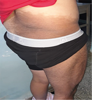
Figure 3: Unilateral linear hyperchromic nevoid lesion in the right middle thoracolumbar region and right thigh.
Complementary exams:
Hemoglobin: 13.8g/L
Leukogram: 9.4 x 10⁹/dL: Polymorph 0.64 Lymphocytes 0.45 Eosinophils: 0.01
Antibody for Hepatitis C Negative
Serology Non-reactive
HIV Negative
He was evaluated by endocrinology who indicated TSH, X-rays of bone age and abdominal ultrasound, all of which were within normal limits.
Pathology: Skin biopsy of lesion: Shows acanthosis with papillomatosis, epidemic hyperkeratosis and hyperpigmentation of the basal layer, scant and focal predominantly lymphocytic inflammatory infiltrate is observed, no evidence of incontinencepigmentaria or melanophages, the histological appearance is suggestive of acanthosis nigricans.Study of the patient is recommended.
Given the complexity of the clinic, the case is collegial with pathological anatomy and eight entities with common clinical, histological and molecular characteristics are considered:
- Epidermal nevi.
- Seborrheic keratosis
- Reticulated and confluent papillomatosis Gougerot and Carteaud
- Unilateral nevoid acanthosis nigricans acronym: RAVEN
- Ichthyosis
- Linear localized epidermal genodermatosis: Porokeratosis, Hailey Hailey disease, Darier disease
- Epidermolytic hyperkeratosis
- Other linear dermatoses: NEVIL
It is decided to carry out a review of the updated bibliography on this type of lesions, reaching the conclusion that it is unilateral nevoid Acanthosis Nigricans with linear disposition.
Nosological diagnosis: Unilateral nevoid acanthosis nigricans.
Behavior: As it is a benign pathology, expectant behavior and follow-up by consultation are decided.
Unilateral nevoid acanthosis nigricans [UNAN] is a recently described entity characterized by lesions, morphologically similar to classic acanthosis nigricans [AN].[1,5] The most prominent features of UNAN include localized distribution, benign course, lack of associations systemic and other tumors, and occurrence due to somatic mosaicism of postzygotic gene mutation.[4,5] It is a dermatosis that mixes clinical aspects of both acanthosis nigricans and epidermal nevus, as shown in the case above.
The remarkable characteristics of the lesions observed distinguish them from other varieties of epidermal nevi [late appearance and velvety surface], as well as from the usual forms of acanthosis nigricans [linear distribution and absence of endocrine or neoplastic context].
In classic AN, the neck is the most recurrently affected site, followed by the axillae, it can also be found on the face, on flexion surfaces, extensor surfaces and intertriginous areas, [1-5] which does not correspond to the case presented. UNAN has a similar morphology to classic AN, but the distribution is unilateral or localized [as occurs in skin lesions that follow Blaschko lines and have an abrupt midline arrest] and manifest during childhood or later. late. [1,3,5] Behavior that was present in the case described above.
Due to the proven benign course of this disease, the treatment generally responds to the aesthetic aspect, [5] so in the case studied it was decided to maintain expectant management and follow-up by consultation.
Unilateral nevoid acanthosis nigricans has a very low incidence worldwide.
Despite the benign course of this disease, its diagnosis is of great importance for the selection of treatment.
Due to the little scientific information available on nevoid acanthosis nigricans, the exposition of the case presented here is of great importance, since it provides the medical community with support material for the study of the behavior of this disease.
- RománSainz, J., SilvestreTorner, N., TabbaraCarrascosa, SS., ImbernónMoya A (2022) Nevoid acanthosisnigricans located on the scalp Dermatol Pract Concept 12. [Crossref].
- Ayaki Matsumoto, KN., Daisuke Tsuruta KS (2021) A Case of Nevoid AcanthosisNigricans SuccessfullyTreated with Topical Ketoconazole Plus Urea. Acta Dermato venerol Croast 29: 167-168. [Crossref].
- ReyesMeza, SE., Guevara Gutiérrez, E., Villanueva Quintero, G., Hernández Arana, S., Tlacuilo Parra (2021) A Nevoid acanthosisnigricans: Report of four cases localized to the umbilicus. Indian J Dermatol Venereol Leprol 87: 660-665. [Crossref].
- Mohammad Ebrahimzadeh, A., Ghanei, N., Shafihosseini, M (2019) Unilateral Nevoid Acanthosis Nigricans on the Back: A Case Report. Acta Medica Iranica 57 :335-337.
- Kumari, I., Kharkar, V (2019) An unusual case of unilateral nevoid acanthosisnigricans with dense lichenoid infiltrate. Indian J Dermatopathol Diagn Dermatol 6: 57-59.