Aims: To compare the visual, anatomic and refractive outcomes in eyes treated with combined phacovitrectomy versus sequential phacoemulsification and vitrectomy.
Methods: A retrospective chart review was performed of eyes with cataract and idiopathic Epiretinal Membrane (ERM) that underwent either combined or sequential phacoemulsification and Pars Plana Vitrectomy (PPV). Data were collected at preoperative (baseline) and Postoperative Month (POM) 1, 3-6, and 12, and the most recent exam beyond POM12 (last exam). Changes from baseline in visual acuity (mean approximate ETDRS letter score), mean Optical Coherence Tomography (OCT) parameters, and mean Refractive Target Error (RTE) calculated as the difference between predicted and actual Spherical Equivalent (SE) were compared between the combined and sequential groups.
Results: Of 100 eyes assessed, 65 had combined phacovitrectomy, 28 had phaco 1st, and 7 had PPV 1st. No significant differences in RTE were observed between groups. No significant differences in mean approximate ETDRS letter score improvement, Central Foveal Thickness (CFT) or Macular Cube Volume (MCV) changes were observed between the combined and either sequential group, or between sequential groups, at any postoperative time point.
Conclusion: Combined and sequential phacoemulsification and vitrectomy procedures produced similar results with respect to visual, anatomic, and refractive outcomes in the population assessed. Surgeons may choose either approach while targeting specific circumstances of each individual patient.
Cataract; Epiretinal Membrane; Phacoemulsification; Phacovitrectomy; Refraction; Vitrectomy
Idiopathic Epiretinal Membrane (ERM) affecting the macula may lead to progressive distortion and impairment of central vision [1]. Pars plana vitrectomy (PPV) with membrane peel is commonly indicated for ERM [2], but is associated with postoperative cataract progression [3] and often requires subsequent Cataract Extraction (CE) [2]. To avoid this or to improve intraoperative view in those with pre-existing cataract [4], combined phacovitrectomy may be performed [5].
Combined phacovitrectomy has been shown to be equally safe [6] and efficacious as separate procedures in terms of restoration of Visual Acuity (VA) and macular anatomy [5,7-9]. The impact of combined procedures on the ability to predict postoperative refraction however, remains unclear. While some prior studies observed a greater tendency toward myopia in individuals who underwent combined phacovitrectomy compared to phacoemulsification alone [10, 11], others did not [12,13]. Few studies have compared Refractive Target Error (RTE) between combined and separate approaches or by the sequence of separate procedures. It is important to understand how the addition and timing of vitrectomy in relation to cataract surgery impacts refractive outcomes in order to improve preoperative planning and limit postoperative deviations from refractive goal.
The purpose of the current study was to compare functional (VA and RTE) and anatomic outcomes (Central Foveal Thickness (CFT) and Macular Cube Volume (MCV) changes) in eyes that underwent combined or separate procedures. We also attempted to compare outcomes stratified by surgical sequence (CE 1st or PPV 1st) in those who had separate procedures.
The study was approved by the Bascom Palmer Eye Institute/University of Miami and Miami Veterans Affairs Hospital Institutional Review Boards. It was conducted in accordance with the principles of the Declaration of Helsinki and was in compliance with the requirements of the United States Health Insurance Portability and Accountability Act.
A retrospective case series was performed of patients at least 18 years of age identified by CPT codes for phacoemulsification combined with or in sequence with PPV and ERM peeling within one year. Patients were excluded from the study if they had intraoperative complications (posterior capsular rupture, vitreous prolapse, dropped nucleus, retinal detachment, choroidal effusion, suprachoroidal hemorrhage), postoperative complications (endophthalmitis, corneal decompensation, non-resolving increased IOP or hypotony, vitreous hemorrhage, optic neuropathy or phototoxicity), were left aphakic during phacoemulsification, or had ocular comorbidities (retinal detachment, vitreous or subretinal hemorrhage, ruptured globe, diabetic retinopathy, or maculopathy secondary to medication use). Children, pregnant women, and prisoners were excluded.
Data Collection
Snellen best-corrected visual acuities (BCVA) were collected from preoperative baseline and at Postoperative Months 1 (POM1), 3-6 (POM3-6), 12 (POM12) records, and the most recent exam beyond POM12 at the time of data collection (last exam). For those that had sequential procedures, BCVA was recorded at baseline and POM1 after the first procedure in addition to the above-mentioned time points for the second procedure. BCVA was converted to approximate ETDRS letter scores for analysis [14]. The same time points were utilized to record Central Foveal Thickness (CFT) and Macular Cube Volume (MCV) from available spectral domain OCT reports (Cirrus, Carl Zeiss Meditec Inc., Dublin, CA or Spectralis, Heidelberg Inc., Germany). The same OCT device was typically used for multiple time points on the same patient. Finally, manifest refraction was recorded at POM1 (after the final procedure) and last exam, and was used to calculate the actual Spherical Equivalent (SE). Axial Length (AL) and target SE based on the SRK/T and Holladay 1 formulas were recorded from IOL Master 500 (Carl Zeiss Meditec, Dublin, CA) biometry, and the Refractive Target Error (RTE) was assessed as the difference between the target (based on the IOL power implanted) and actual postoperative SE. The RTE was calculated for both the SRK/T and Holladay 1 formulas for each eye, as available in the charts. The surgeon typically targeted a SE of plano or mild myopia. The data were collected over the last 5 years, as the transition to electronic health record at the Bascom Palmer Eye Institute occurred, thus some data that had not been scanned into the electronic system were not available.
Statistical Analysis
VA, OCT measurements, and RTE values at each postoperative time point were compared to baseline values of these same patients for the following: postoperative months 1 (POM1), 3-6 (POM3-6), 12 (POM12), and the most recent exam beyond POM12 (last exam). Statistical analysis was performed using SAS Version 9.4 (Cary, NC, USA). Visits were classified into follow-up examination windows, defined as: 1 month (3-6 weeks); 3 -6 months (2-7 months), and 12 months (9-13 months). If more than one visit occurred within a follow-up window, the one closest to the defined interval visit was selected for analysis. For the purpose of calculating differences in visual acuity, Snellen visual acuities were converted into approximate Early Treatment Diabetic Retinopathy Study (approx ETDRS) letter scores as previously described [14]. Between-group differences in categorical variables such as gender and laterality were assessed using the chi-square test or Fisher’s Exact test, when the assumptions of the chi-square test were invalid. Continuous variables such as axial length, VA, OCT measurements, spherical equivalence, and RTE were assessed for normal distributions using the Shapiro-Wilk and Kolmogorov-Smirnov tests. Because normal distributions could not be assumed for most continuous variables, between-group differences were assessed using the paired-sample Wilcoxon signed-rank non-parametric test. A p-value of 0.05 or less was considered statistically significant.
Demographics
Demographic data are presented in detail in Table 1. One hundred eyes were included, with sixty-five of these undergoing combined phacovitrectomy procedures (Combo group). The mean age of the Combo group was 67±10.8 years and the mean AL was 24.3±1.3mm. Thirty-five eyes had separate procedures. Of these, 28 (80%) had phacoemulsification first (CE 1st). The mean age of the CE 1st group was 66.4±9.4 years, mean AL was 24.6±2.1mm, and the mean interval between procedures was 141.4±92.8 days. Seven eyes had PPV 1st. The mean age in the PPV 1st group was 65±4.2 years, mean AL was 24.7±1.7mm, and the mean interval between procedures was 206.7±91.4 days. No significant differences in baseline characteristics were observed between the three groups. (Table 1)
Table 1. Demographic Data for Combined and Sequential Groups.
| |
Combined phacovitrectomy |
Sequential: CE 1st, PPV 2nd |
Sequential: PPV 1st, CE 2nd |
P-value* |
Number of patients |
65 (65%) |
28 (28%) |
7 (7%) |
|
Mean (SD) Age (years) |
67 (10.8) |
66.4 (9.4) |
65 (4.2) |
0.4186, 0.7123, 0.7100, 0.5291 |
% Female |
26.2 |
25 |
28.6 |
1.000, 0.9071, 1.000, 0.9619 |
Laterality (% OD) |
52.3 |
64.3 |
57.1 |
1.000, 0.2859, 1.000, 0.3107 |
Mean (SD) Axial Length (mm) |
24.3 (1.3) |
24.6 (2.1) |
24.7 (1.7) |
0.4690, 0.5645, 0.4560, 0.4428 |
Mean (SD) days between procedures |
N/A |
141.4 (92.8) |
206.7 (91.4) |
N/A, N/A, 0.0869, N/A |
Mean (SD) days since last procedure |
722.2 (626.9) |
805.9 (369.7) |
560.7 (332.7) |
0.7991, 0.2388, 0.1220, 0.3598 |
*Sequence of P-values displayed = Combined versus PPV 1st, Combined versus CE 1st, CE 1st versus PPV 1st, Combined versus All Staged
N/A = not applicable
Visual Acuity
There were no significant differences in VA between the groups at baseline.
No significant differences were observed between the Combo and either sequential groups in VA improvement from baseline to any postoperative time point, or the percentage of individuals demonstrating at least 3 lines of improvement or 3-line loss. Similarly, no significant differences were observed in any of these parameters between either of the two sequential groups. Median Snellen VA consistently improved to at least 20/50 by last exam and mean approximate ETDRS letter scores increased from baseline to last exam (mean of 14 to 18 letters) in all three groups. (Figure 1, Table 2) Forty to over fifty percent of cases improved by at least 3 lines by 12 postoperative months in all groups. (Table 2)
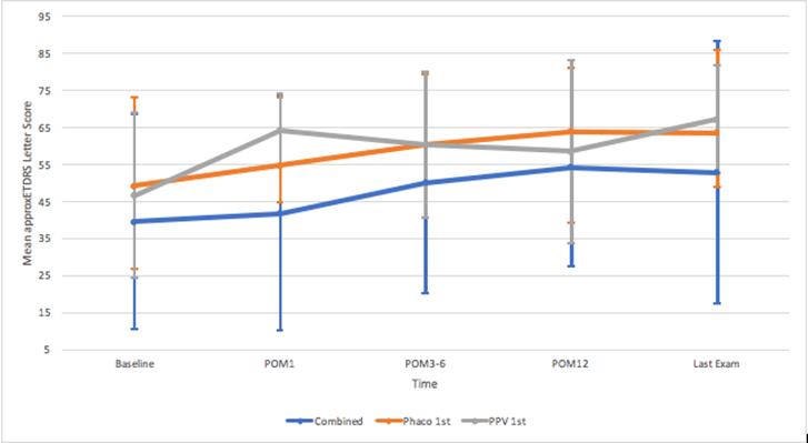
Figure 1. Mean approximate ETDRS Letter Scores from Baseline to Last Exam for Patients in Combined and Sequential Groups.
*For sequential groups, POM1 is after the second procedure.
POM=postoperative month
Table 2. Visual Acuity Measurements at Baseline and Through Last Postoperative Exam for Patients in Combined and Sequential Groups.
Time Point |
Parameter* |
Combined phacovitrectomy |
Sequential: CE 1st, PPV 2nd |
Sequential: PPV 1st, CE 2nd |
P-Value |
Combined versus Sequential CE 1st |
Combined versus Sequential PPV 1st |
Sequential CE 1st versus PPV 1st |
Combined versus All Sequential |
Baseline |
Snellen Median VA |
20/100 |
20/70 |
20/150 |
|
|
|
|
Snellen [range] |
[20/30, LP] |
[20/25, CF] |
[20/20,20/400] |
|
|
|
|
approxETDRS Mean±SD |
40±29.0 |
49±24.0 |
47±22.4 |
0.1998 |
0.9829 |
0.452 |
0.2582 |
approxETDRS Median[range] |
50[-39, 76] |
58[-15,80] |
41[20, 85] |
|
|
|
|
N eyes |
N=63 |
N=26 |
N=6 |
|
|
|
|
POM1 |
Snellen Median VA |
20/100 |
20/60 |
20/50+3 |
|
|
|
|
Snellen [range] |
[20/20, LP] |
[20/25,20/400] |
[20/30,20/100] |
|
|
|
|
approxETDRS mean letters improved±SD from baseline |
3±26.3 |
5.5±22.3 |
17.7±21.9 |
0.7773 |
0.2188 |
0.3334 |
0.4824 |
3-line improvement: N (%) |
18 (30.0) |
7 (26.9) |
3 (50) |
0.7729 |
0.3732 |
0.3461 |
0.9012 |
3-line loss: N (%) |
12 (20.0) |
5 (19.2) |
0 (0.0) |
0.9344 |
0.5823 |
0.5546 |
0.6066 |
N eyes |
N=60 |
N=26 |
N=6 |
|
|
|
|
POM3-6 |
Snellen Median VA |
20/63 |
20/60 |
20/50+1 |
|
|
|
|
Snellen [range] |
[20/20, LP] |
[20/20,20/800] |
[20/30,20/200] |
|
|
|
|
approxETDRS mean letters improved±SD from baseline |
10.1±24.3 |
11.6±18.5 |
22.0±31.2 |
0.7394 |
0.7237 |
0.8492 |
0.852 |
3-line improvement: N (%) |
27 (50.0) |
9 (36.0) |
2 (50.0) |
0.2452 |
1 |
0.6221 |
0.2927 |
3-line loss: N (%) |
7 (13.0) |
1 (4.0) |
0 (0.0) |
0.4242 |
1 |
1 |
0.2507 |
N eyes |
N=54 |
N=25 |
N=4 |
|
|
|
|
POM12 |
Snellen Median VA |
20/50 |
20/50 |
20/70+1 |
|
|
|
|
Snellen [range] |
[20/20, HM] |
[20/20,20/300] |
[20/30,20/150] |
|
|
|
|
Approx. ETDRS mean letters improved±SD from baseline |
14.0±22.9 |
16.4±27.7 |
28.0±39.6 |
0.8273 |
0.7923 |
0.8021 |
0.7855 |
3-line improvement: N (%) |
16 (44.4) |
10 (43.5) |
1 (50.0) |
0.9419 |
1 |
1 |
0.9726 |
3-line loss: N (%) |
3 (8.3) |
3 (13.0) |
0 (0.0) |
1 |
1 |
0.5594 |
0.6363 |
N eyes |
N=36 |
N=23 |
N=2 |
|
|
|
|
Last Exam |
Snellen Median VA |
20/50 |
20/40 |
20/40 |
|
|
|
|
Snellen [range] |
[20/20, LP] |
[20/20, CF] |
[20/20,20/100] |
|
|
|
|
approxETDRS mean letters improved+SD from baseline |
14.0±26.6 |
14.3±29.2 |
18.2±24.6 |
0.7813 |
0.9508 |
0.8931 |
0.8242 |
3-line improvement: N (%) |
35 (58.3) |
11 (42.3) |
2 (40.0) |
0.1712 |
0.6444 |
1 |
0.1376 |
3-line loss: N (%) |
6 (10.0) |
3 (11.5) |
0 (0.0) |
1 |
1 |
1 |
1 |
N eyes |
N=60 |
N=26 |
N=5 |
|
|
|
|
*Parameters at each postoperative visit were compared to the baseline values of those patients only.
At each time point, only eyes that had available data at both baseline and the follow-up time point were included in the analysis.
Abbreviations: ApproxETDRS=approximate ETDRS letters
Macular Anatomy
There were no significant differences in mean CFT or MCV at baseline or any postoperative time point between the groups (Table 3). Mean CFT tended to decrease with postoperative time in all three groups, and reached minimal thickness at POM12. (Figure 2)
Table 3. SD-OCT Central Foveal Thickness (CFT) and Macular Cube Volume (MCV) Measurements at Baseline and Through Last Postoperative Exam in Combined and Sequential Groups.
Time Point |
Parameter |
Combined phacovitrectomy |
Sequential: CE 1st, PPV 2nd |
Sequential: PPV 1st, CE 2nd |
P-Value |
Combined versus Sequential CE 1st |
Combined versus Sequential PPV 1st |
Sequential CE 1st versus PPV 1st |
Combined versus All Sequential |
Baseline |
Mean CFT (µm) ±SD |
482.4±141.0 |
478.3±212.5 |
412.3±142.2 |
0.2757 |
0.4976 |
0.694 |
0.2873 |
Median CFT [range] |
468 [225, 819] |
440 [168, 998] |
442 [202, 608] |
N eyes |
N=55 |
N=18 |
N=7 |
Mean MCV (mm3) ±SD |
12±2.95 |
11.34±3.17 |
12.39±2.85 |
0.7809 |
0.6868 |
0.9036 |
0.8477 |
Median MCV [range] |
12 [2, 20] |
12 [3, 17] |
13 [10, 18] |
N eyes |
N=55 |
N=18 |
N=7 |
POM1 |
Mean CFT ±SD |
405.4±113.8 |
429.4±142.5 |
339.7±53.8 |
|
|
|
|
Median CFT [range] |
414 [183, 711] |
420 [216, 766] |
362 [272, 393] |
|
|
|
|
Mean change ±SD from baseline |
-95.7±157.1 |
-91.0±167.9 |
-107.7±93.9 |
0.6583 |
0.756 |
0.4596 |
0.9715 |
N eyes |
N=43 |
N=15 |
N=6 |
|
|
|
|
Mean MCV ±SD |
11.36±1.91 |
11.33±1.99 |
11.38±1.23 |
|
|
|
|
Median MCV [range] |
12 [6, 16] |
11 [6, 14] |
11 [10, 14] |
|
|
|
|
Mean change ±SD from baseline |
-1.01±2.60 |
-0.20±2.71 |
-1.42±2.48 |
0.6148 |
0.7831 |
0.7554 |
0.6421 |
N eyes |
N=43 |
N=15 |
N=6 |
|
|
|
|
POM3-6 |
Mean CFT ±SD |
381.2±111.2 |
385.1±151.3 |
367.0±N/A |
|
|
|
|
Median CFT [range] |
361 [146, 670] |
368 [151, 785] |
367 [367, 367] |
|
|
|
|
Mean change ±SD from baseline |
-105.6±132.8 |
-99.0±190.1 |
-119.0±N/A |
0.7875 |
0.8218 |
0.8472 |
0.8767 |
N eyes |
N=44 |
N=17 |
N=1 |
|
|
|
|
Mean MCV ±SD |
11.27±2.39 |
11.02±2.10 |
11.60±N/A |
|
|
|
|
Median MCV [range] |
11 [7, 22] |
11 [6, 16] |
12 [12, 12] |
|
|
|
|
Mean change ±SD from baseline |
-0.75±2.80 |
-0.26±2.36 |
-5.90±N/A |
0.1142 |
0.6407 |
0.1231 |
0.9012 |
N eyes |
N=44 |
N=17 |
N=1 |
|
|
|
|
POM12 |
Mean CFT ±SD |
360.1±109.2 |
333.3±141.4 |
366.0±N/A |
|
|
|
|
Median CFT [range] |
352 [131, 573] |
322 [99, 766] |
366 [366, 366] |
|
|
|
|
Mean change ±SD from baseline |
-126.5±142.7 |
-158.8±170.4 |
-242.0±N/A |
0.37 |
0.6084 |
0.358 |
0.4971 |
N eyes |
N=28 |
N=16 |
N=1 |
|
|
|
|
Mean MCV ±SD |
11.02±1.49 |
10.37±2.21 |
13.5±N/A |
|
|
|
|
Median MCV [range] |
11 [8, 14] |
10 [6, 15] |
14 [14, 14] |
|
|
|
|
Mean change ±SD from baseline |
-1.31±1.95 |
-0.90±2.57 |
-0.10±N/A |
0.6755 |
0.9222 |
0.358 |
1 |
N eyes |
N=28 |
N=16 |
N=1 |
|
|
|
|
Last Exam |
Mean CFT ±SD |
367.1±118.2 |
335.5±139.7 |
339.8±56.4 |
|
|
|
|
Median CFT [range] |
355 [116, 670] |
320 [99, 739] |
350 [272, 399] |
|
|
|
|
Mean change ±SD from baseline |
-123.8±130.1 |
-148.6±192.3 |
-107.5±115.4 |
0.9412 |
0.7549 |
1 |
0.8301 |
N eyes |
N=35 |
N=17 |
N=6 |
|
|
|
|
Mean MCV ±SD |
10.88±1.63 |
10.21±2.30 |
11.13±1.06 |
|
|
|
|
Median MCV [range] |
11 [8, 15] |
11 [5, 15] |
11 [10, 13] |
|
|
|
|
Mean change ±SD from baseline |
-1.49±1.91 |
-1.07±2.12 |
-1.67±2.48 |
1 |
0.8453 |
0.9163 |
0.8799 |
N eyes |
N=35 |
N=17 |
N=6 |
|
|
|
|
At each time point, only eyes that had available data at both baseline and the follow-up time point were included in the analysis.
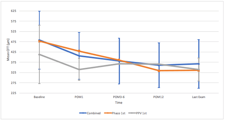
Figure 2. SD-OCT Mean Central Foveal Thickness (CFT) Measurements from Baseline to Last Exam for Patients in Combined and Sequential Groups.
*For sequential groups, POM1 is after the second procedure.
POM=postoperative month
Refractive Target Error
Regardless of the formula used to calculate target SE (SRK/T or Holladay 1), the mean RTE was between -0.64D and +0.19D at all postoperative time points for all three groups (Combo, CE 1st, PPV 1st). The only significant difference was found at POM1 when comparing the mean RTE for the SRK/T formula alone for combined cases versus all sequential cases. The mean RTE for the SRK/T formula at POM1 was significantly more myopic in the Combo group (-0.64±1.33D) compared to those who underwent separate procedures (0.17±1.27D for CE 1st and 0.081±0.71D for PPV 1st, P=0.0316). No other significant differences in mean RTE were observed between the combined and either sequential group, or between sequential groups. (Table 4)
When averaging target SE from both SRK/T and Holladay 1 formulas, the mean RTE was slightly more myopic for the Combo group at POM1 (-0.47±1.26D) and last exam (-0.21±1.37D) compared to the sequential groups (which tended to be slightly hyperopic), however the differences were not significant. (Table 4)
Table 4. Mean Target SE, Mean Actual SE, and Mean Refractive Target Error (RTE) for Patients in Combined and Sequential Groups.
Formula |
Time Point |
Parameter (D) |
Combined phacovitrectomy |
Sequential: CE 1st, PPV 2nd |
Sequential: PPV 1st, CE 2nd |
P-Value |
Combined versus Sequential CE 1st |
Combined versus Sequential PPV 1st |
Sequential CE 1st versus PPV 1st |
Combined versus All Sequential |
SRK/T |
POM1 |
Mean Target SE±SD |
-0.17±0.56 |
-0.56±1.08 |
-0.46±0.14 |
0.9608 |
0.3628 |
0.1262 |
0.6991 |
Mean Actual SE±SD |
-0.81±1.35 |
-0.39±1.36 |
-0.38±0.61 |
0.2583 |
0.4594 |
1 |
0.2105 |
Mean RTE±SD |
-0.64±1.33 |
0.17±1.27 |
0.081±0.71 |
0.0525 |
0.1976 |
0.9188 |
0.0316* |
N eyes |
N=38 |
N=20 |
N=5 |
|
|
|
|
Last Exam |
Mean Target SE±SD |
-0.16±0.53 |
-0.53±1.06 |
-0.99±0.89 |
0.7585 |
0.0951 |
0.0885 |
0.4364 |
Mean Actual SE±SD |
-0.41±1.45 |
-0.35±1.67 |
-1.00±0.78 |
0.8077 |
0.3268 |
0.3126 |
0.6049 |
Mean RTE±SD |
-0.26±1.38 |
0.19±1.68 |
-0.01±0.75 |
0.497 |
0.4629 |
0.7934 |
0.399 |
N eyes |
N=43 |
N=21 |
N=3 |
|
|
|
|
Holladay 1 |
POM1 |
Mean Target SE±SD |
-0.30±0.43 |
-0.60±0.98 |
-0.49±0.13 |
0.8173 |
0.2031 |
0.26 |
0.5176 |
Mean Actual SE±SD |
-0.72±1.28 |
-0.44±1.25 |
-0.38±0.61 |
0.588 |
0.6188 |
0.862 |
0.5135 |
Mean RTE±SD |
-0.42±1.29 |
0.16±1.16 |
0.113±0.72 |
0.2218 |
0.3517 |
0.8399 |
0.1613 |
N eyes |
N=47 |
N=24 |
N=5 |
|
|
|
|
Last Exam |
Mean Target SE±SD |
-0.28±0.42 |
-0.59±0.96 |
-0.92±0.77 |
0.5994 |
0.0892 |
0.1473 |
0.3404 |
Mean Actual SE±SD |
-0.41±1.44 |
-0.40±1.54 |
-1.00±0.78 |
0.765 |
0.3163 |
0.3131 |
0.5794 |
Mean RTE±SD |
-0.133±1.34 |
0.19±1.57 |
-0.083±0.69 |
0.6384 |
0.6504 |
0.8527 |
0.5729 |
N eyes |
N=51 |
N=25 |
N=3 |
|
|
|
|
Mean of SRK/T and Holladay 1† |
POM1 |
Mean RTE±SD |
-0.47±1.26 |
0.12±1.18 |
0.10±0.72 |
0.175 |
0.2827 |
0.795 |
0.1161 |
Last Exam |
Mean RTE±SD |
-0.21±1.37 |
0.15±1.57 |
-0.05±0.72 |
0.5631 |
0.4447 |
0.7663 |
0.4534 |
*=P<0.05
†= If there was only data for a single formula (SRK/T or Holladay) then this is simply the mean RTE for that formula alone.
At each time point, only eyes that had available data at both baseline and the follow-up time point were included in the analysis.
As expected, based on prior studies [5,7-9], we observed no significant differences in postoperative improvement in vision and restoration of macular anatomy between those that underwent combined and sequential procedures. All three study groups had mean refractive outcomes well within ±0.5D of target, consistent with previous reporting [15].
The Combo group had significantly more myopic RTE compared to sequential cases only at POM1 if SRK/T formula was used. However, the Combo group was not significantly different from sequential groups despite having slightly more myopic mean RTE compared to either sequential group at POM1 or the last exam if Holladay 1 or its mean with SRK/T was used to predict SE. No other RTE differences achieved statistical significance at any other time point. Thus, the surgeon may use either formula while choosing IOL for these cases. This is in general agreement with the findings of Hamoudi et. al., who reported no significant differences in RTE at POM12 between those who had combined and separate procedures, regardless of sequence [17]. They described a slight myopic outcome, which is generally desirable, in those who had combined phacovitrectomy (-0.15±0.61D) and in those who had separate procedures with phacoemulsification (-0.18±0.85D) or PPV first (-0.19±0.83D).
The preferred approach to patients with cataract and epiretinal membrane varies among surgeons. Combined phacovitrectomy has the advantages of being more cost effective17 and some reported lower surgical risk with this approach. For example, a retrospective review of 54 eyes documented significantly lower rates of posterior capsular rupture in those who had combined (4.5%) compared to sequential procedures (11.4%) (P=0.049) [18]. Theoretically, undergoing a single surgery limits the endophthalmitis risk compared to having multiple procedures. The combined approach also eliminates the risk for postoperative cataract progression, which is commonly encountered after isolated PPV and often requires subsequent CE [3]. Decreased vitreous and zonular support after isolated PPV may make it more difficult to perform subsequent CE. Also, improved intraoperative view by initial CE in the combined approach may enable more thorough removal of anterior vitreous compared to an isolated PPV. Despite these advantages, phacovitrectomy may be associated with greater postoperative RTE than cataract surgery alone [10,11]. This is possibly due to vitrectomy-associated changes in refractive indices and parameters that influence IOL power calculation, such as AL and Anterior Chamber Depth (ACD) [11].
Increased postoperative AL after ERM peeling may contribute to myopic shift. This may occur when retinal thickening due to the presence of ERM impacts the reliability of ultrasonographic IOL calculations, or secondary to scleral thinning at sclerotomy sites used in PPV [11]. Despite these possibilities, Kim et. al. reported no significant changes in AL measured by IOL Master (Carl Zeiss Meditec, Dublin, CA) after combined phacovitrectomy [11]. They attributed this to the fact that optical biometry measures down to the Retinal Pigment Epithelium (RPE) and should not be influenced by differential retinal thickness [11]. While we also relied on IOL Master 500 for power calculations, we did not evaluate for the possibility that reflection of the optical signal off a dense ERM could alter AL measurement and ultimately contribute to the observed slight myopic shift [16].
ACD is considered to be a reliable marker of Effective Intraocular Lens Position (ELP), which is an important factor in IOL power calculations [19]. A 1mm forward movement of lens implant causes -1.5D of myopia [16], and previous studies have reported significantly increased ACD in patients who underwent combined phacovitrectomy compared to phacoemulsification alone [10]. It is possible that the differential changes in ACD and IOL position contributed to the slight differences in RTE that we observed between surgical groups.
Combined phacovitrectomy is not without disadvantages. Phacoemulsification may result in corneal hydration and a more difficult view for the retinal surgeon. If the cataract is removed after vitrectomy is performed, decreased scleral rigidity may make extraction more difficult, in our experience. A lengthy cataract extraction may increase the risk for intraoperative corneal decompensation or increased inflammation and subsequent macular edema compared to separate procedures. Our observed trend of decreasing CFT with postoperative time likely represents the resolution of macular edema in the population studied.
The findings must be considered in light of the data limitations. First, multiple surgeons performed the procedures, which may confound outcomes due to inconsistent techniques and IOL choices and calculations. Second, many of our patients were elderly male veterans, thus future studies would need to substantiate the findings in more diverse populations. Third, our sample size was relatively small, especially in the PPV1st group. The conclusions of the paper should be considered in light of this limitation, which diminished the power of our statistical analysis. Future studies are needed to explore this topic in a larger cohort.
Despite these limitations, our findings of equal visual and anatomic outcomes are encouraging as they free the surgeon to choose the best surgical approach for each individual patient. Since the sequence of surgery does not appear to significantly impact the outcomes in the population studied, the surgeon may choose either combined or sequential approach based on the severity of the cataract and epiretinal membrane.
Data analysis for this work was supported by NIH Center Core Grant P30EY014801, Research to Prevent Blindness Unrestricted Grant. The sponsor or funding organization had no role in the design and conduct of the study; collection or interpretation of the data, and preparation or approval of the manuscript. None of the authors has conflicts of interest pertaining to this work. Contents of this article were presented at the 2019 Association for Research in Vision and Ophthalmology (ARVO) conference in Vancouver, BC on 4/28/19.
- Bu SC, Kuijer R, Li XR, Hooymans JM, Los LI (2014) Idiopathic epiretinal membrane. Retina. 34:2317-2335. [Crossref]
- Jackson TL, Donachie PH, Sparrow JM, Johnston RL (2013) United Kingdom National Ophthalmology Database Study of Vitreoretinal Surgery: report 1; case mix, complications, and cataract. Eye (Lond). 27:644-651. [Crossref]
- Feng H, Adelman RA (2014) Cataract formation following vitreoretinal procedures. Clin Ophthalmol. 8:1957-1965. [Crossref]
- Demetriades AM, Gottsch JD, Thomsen R, Azab A, Stark WJ, et al. (2003) Combined phacoemulsification, intraocular lens implantation, and vitrectomy for eyes with coexisting cataract and vitreoretinal pathology. Am J Ophthalmol. 135:291-296. [Crossref]
- Wu Z, Zhang J, Chen Y, Gao R, Lin Z (2012) Efficacy of phacovitrectomy combined with internal limiting membrane peeling for macular diseases. Eye Sci. 27:25-29. [Crossref]
- Savastano A, Savastano MC, Barca F, Petrarchini F, Mariotti C, et al. (2014) Combining cataract surgery with 25-gauge high-speed pars plana vitrectomy: results from a retrospective study. Ophthalmology. 121:299-304. [Crossref]
- Dugas B, Ouled-Moussa R, Lafontaine PO, Guillaubey A, Berrod JP, et al. (2010) Idiopathic epiretinal macular membrane and cataract extraction: combined versus consecutive surgery. Am J Ophthalmol. 149:302-306. [Crossref]
- Yiu G, Marra KV, Wagley S, Krishnan S, Sandhu H, et al. (2013) Surgical outcomes after epiretinal membrane peeling combined with cataract surgery. Br J Ophthalmol. 97:1197-1201. [Crossref]
- Kauffmann Y, Ramel JC, Isaico R, De Lazzer A, Bron AM, et al. (2017) Long-Term Anatomical and Functional Outcomes after Combined Cataract and Idiopathic Epiretinal Membrane Surgery. Ophthalmic Res. 57:125-134. [Crossref]
- Falkner-Radler CI, Benesch T, Binder S (2008) Accuracy of preoperative biometry in vitrectomy combined with cataract surgery for patients with epiretinal membranes and macular holes: results of a prospective controlled clinical trial. J Cataract Refract Surg. 34:1754-1760. [Crossref]
- Kim M, Kim HE, Lee DH, Koh HJ, Lee SC, et al. (2015) Intraocular lens power estimation in combined phacoemulsification and pars plana vitrectomy in eyes with epiretinal membranes: a case-control study. Yonsei Med J. 56:805-811. [Crossref]
- Shi L, Chang JS, Suh LH, Chang S (2019) Differences in Refractive Outcomes between Phacoemulsification for Cataract Alone and Combined Phacoemulsification and Vitrectomy for Epiretinal Membrane. Retina. 39:1410-1415. [Crossref]
- van der Geest LJ, Siemerink MJ, Mura M, Mourits MP, Lapid-Gortzak R (2016) Refractive outcomes after phacovitrectomy surgery. J Cataract Refract Surg. 42:840-845. [Crossref]
- Gregori NZ, Feuer W, Rosenfeld PJ (2010) Novel method for analyzing snellen visual acuity measurements. Retina. 30:1046-1050. [Crossref]
- Ercan ZE, Akkoyun I, Yaman Pinarci E, Yilmaz G, Topcu H (2017) Refractive outcome comparison between vitreomacular interface disorders after phacovitrectomy. J Cataract Refract Surg. 43:1068-1071. [Crossref]
- Hamoudi H, Correll Christensen U, La Cour M (2018) Epiretinal membrane surgery: an analysis of 2-step sequential- or combined phacovitrectomy surgery on refraction and macular anatomy in a prospective trial. Acta Ophthalmol. 96:243-250. [Crossref]
- Seider MI, Michael Lahey J, Fellenbaum PS (2014) Cost of phacovitrectomy versus vitrectomy and sequential phacoemulsification. Retina. 34:1112-1115. [Crossref]
- Lee JY, Kim KH, Shin KH, Han DH, Lee DY, et al. (2012) Comparison of intraoperative complications of phacoemulsification between sequential and combined procedures of pars plana vitrectomy and cataract surgery. Retina. 32:2026-2033. [Crossref]
- Olsen T (2007) Improved accuracy of intraocular lens power calculation with the Zeiss IOLMaster. Acta Ophthalmol Scand. 85:84-87. [Crossref]