Background: Adolescent idiopathic scoliosis (AIS) results in three-dimensional malrotation that cannot be fully characterized on 2-view radiographs. Pedicle screw placement is the preferred method to safely and effectively reduce axial rotation, and screw placement at the scoliotic apex has been considered paramount.
Purpose: To evaluate axial rotation correction on CT in AIS patients undergoing pedicle screw placement at the thoracic curve apex. To evaluate whether proximity and number of pedicle screws near the curve apex correlates with improved correction of axial rotation.
Methods: Retrospective review was made of patients <18 years with AIS and primary thoracic curvature who underwent surgery and had preoperative and postoperative CTs. During a 17-month interval, 41 patients were included (34 F/7M) between the ages of 12 and 17 (average=14.2) years. Operations were performed at single institution by single pediatric orthopedic surgeon. Average correction in axial rotation on CT was calculated using the Ho method. Spearman correlation coefficient was calculated to assess the relationship between reduction in axial rotation and the number of screws concentrated near the apex or total number of screws.
Results: Average change in axial rotation after pedicle screw placement was 5 degrees (38% change). No statistically significant correlation was found between change in axial rotation and number of screws at the apex, location of closest screw located above or below the apex, and number of screws located above or below the apex.
Conclusion: If pedicle instrumentation at the scoliotic apex does not contribute to axial correction then this level could be avoided, thereby decreasing costs and complications related to screw breech.
Level of Evidence: Therapeutic Study - Level III
Adolescent Idiopathic Scoliosis; Computed Tomography; Pedicle Screws; Postoperative; Axial Rotation
Diagnosis, prognosis, and selection of treatment options for patients with Adolescent Idiopathic Scoliosis (AIS) has historically been reliant on radiographic evaluation of Cobb angle in the coronal and sagittal planes. A better understanding of scoliosis over the past few decades has demonstrated that scoliosis is a three-dimensional deformity [1-3]. Recent focus has been placed on the evaluation and correction of the third dimension (axial rotation), which contributes to trunk rotation and cosmetic deformity [1]. This component of the scoliotic curve cannot be adequately assessed from 2-view radiographs [1,4]. Many authors have identified the apex of the scoliotic curve as the focus of maximum rotation [1,5,6]. The thoracic vertebral bodies near the apex are also known to be the site of the most hypoplastic and dysplastic pedicles in AIS patients [7,8]. The current preferred method of spinal correction in AIS is the pedicle screw construct which requires precise positioning of pedicle screws in order to avoid damage to adjacent nervous and vascular structures.
Computed Tomography (CT) has been shown to be the most accurate way to evaluate the spine both pre- and postoperatively and has been shown to be better than radiography at evaluating axial rotation [1,9,10]. As the maximal axial rotation in AIS patients involves the vertebral levels with the most hypo plastic and distorted pedicles, the concern for complications related to placement of pedicle screws at or near the thoracic apex was the impetus for this study. This study was designed to evaluate whether pedicle screw fixation contributed to postoperative improvement in axial rotation, and whether the total number of screws placed (screw density) was associated with improved degrees of correction in axial rotation. To our knowledge, this is the first study to evaluate the contribution of screw position (relative to the curve apex) and screw density to the overall correction of axial rotation in AIS patients using CT imaging.
Approval to perform a retrospective study was obtained from our Institutional Review Board.
Patients
We conducted a retrospective review of patients less than 18 years of age with a diagnosis of adolescent idiopathic scoliosis and primary thoracic curvature who had undergone corrective surgery as well as preoperative and postoperative CT scans. A total of 41 patients who underwent surgery between July 2012 and December 2013 were included. (34 F/7 M) between the ages of 12 and 17 (average=14.2) years. All operative procedures were performed at a single institution by a single pediatric orthopedic surgeon.
CT Interpretation
All CT examinations were interpreted independently by two M.D. candidates and reviewed by a senior pediatric radiologist. All CT examinations were performed on a GE VCT 64 (GE Healthcare, Chicago, USA), Philips Brilliance 64 (Philips, Eindhoven, NL), or Philips Ingenuity 128 scanner (Philips, Eindhoven, NL). Preoperative imaging consisted of axial 0.625 mm or 0.8 mm images with 1.5 mm coronal and sagittal reconstructions. Postoperative imaging consisted of axial 2.5 mm images with 1.0 mm coronal and sagittal reconstructions. Number and placement of pedicle screw fixation were documented relative to the scoliotic apex, and axial rotation of the apical vertebra was measured on pre- and postoperative CT scans.
Correction in axial rotation achieved by spinal fusion
The apical vertebra of the thoracic curvature was defined as the vertebra at the point of maximal curvature, and was determined using the coronal reconstruction on preoperative CT. Using axial CT images and applying the Ho method [9], axial rotation of the apical vertebra was calculated. Subsequently, the identical apical vertebra was examined, and axial rotation calculated from the axial CT image obtained post-surgically. Preoperative and preoperative values were compared.
orrection in axial rotation relative to proximity of pedicle screws to the curve apex
The number of screws placed in the apical vertebra was recorded. The number of screws placed in the three vertebrae central to the apex (defined as the apical vertebra and the two flanking vertebrae) were recorded. The number of screws placed at the curve apex and within the three vertebral bodies central to the apex were compared to the total change and percentage change in axial rotation.
Correction in axial rotation relative to the total number of pedicle screws placed
The total number of screws placed per patient was recorded and correlated with the total change and percentage change in axial rotation.
Statistical analysis
Statistical analysis was performed by our institutional statistician utilizing IBM® SPSS® Statistics version 21 software. The Spearman correlation coefficient was selected due to the limited variability of the apex value. Although linear regression and correlation is typically used to determine the relationship between two variables, these analyses assume a normal distribution of data. In our study, there was limited variability in the number of screws placed at, above, or below the apex (0, 1, or 2). Because these values were not normally distributed, another method was required to determine the relationship between screws placed at apex, just above the apex, or just below the apex and the resultant decrease in axial rotation. The Spearman correlation coefficient does not assume a normal distribution of any variables and was thus used for data analysis.
Correction in axial rotation achieved by spinal fusion
Using axial CT images and applying the Ho method, axial rotation of the apical vertebra was determined and compared pre and postoperatively. The average change in axial rotation is 5 degrees or 38% change between the preoperative and postoperative exam.
Correction in axial rotation relative to the total number of pedicle screws placed
The total number of patients studied was 41. The average number of screws placed per patient was 14.2 (range 10 to 23) with a standard deviation of 3.1 (Table 1). It was determined that no statistically significant relationship existed between the change or percent change in axial rotation and the total number of screws used in fusion (p = 0.59 and 0.65) (Table 2).
Table 1. Scatterplot diagram shows change in axial rotation based upon total number of screws in 41 patients.
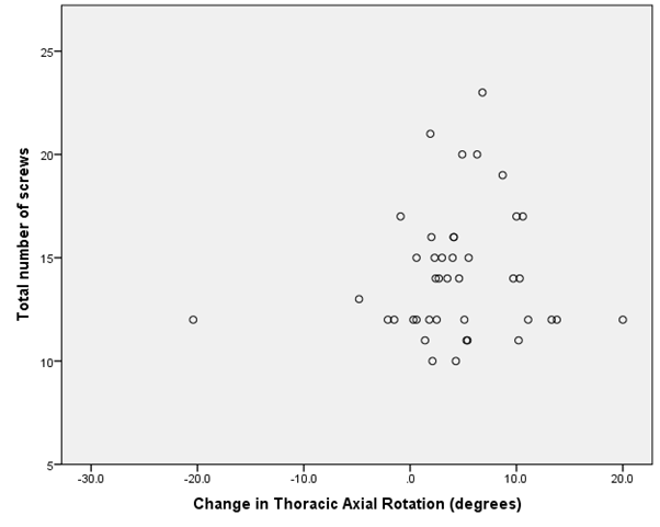
Table 2. Spearman correlation coefficient demonstrates no statistically significant relationship between change in axial rotation and total number of screws.
|
Correlations |
|
|
Change |
% Change |
Total number of screws |
|
Spearman's rho |
Change |
Correlation coefficient |
1.000 |
0.858** |
0.089 |
Sig. (2-tailed) |
- |
0.000 |
0.582 |
n |
41 |
41 |
41 |
|
Percent change |
Correlation coefficient |
0.858** |
1.000 |
0.073 |
Sig. (2-tailed) |
.000 |
- |
0.651 |
n |
41 |
41 |
41 |
|
Total number of screws |
Correlation coefficient |
0.089 |
0.073 |
1.000 |
Sig. (2-tailed) |
0.582 |
0.651 |
- |
n |
41 |
41 |
41 |
**. Correlation is significant at the 0.01 level (2-tailed).
Correction in axial rotation relative to proximity of pedicle screws to the curve apex
The average number of screws placed at the curve apex was 1.7 with 71% (29/41) of patients having 2 screws, 24% (10/41) having one screw, and 5% (2/41) having no screws placed at the apex (Table 3). There was no statistical correlation between the number of screws placed at the level of the apical curvature and the degree or percentage of axial rotation correction (p = 0.78 and 0.99) (Table 4). Of the total number of patients, 81% (33/41) had instrumentation of the three levels central to the thoracic apex. Each of these patients had pedicle screws placed at the apical vertebra and within the immediately adjacent superior and inferior vertebra. The majority of patients (73%, 24/33) had 2 screws placed at all three levels (a total of 6 of screws within the 3 vertebral bodies). Six per cent (2/33) had 5 screws, 15% (5/33) had 4 screws and 6% (2/33) had 3 screws. Evaluation of this group identified no statistically significant relationship between the change in axial rotation and the number of screws placed within the three vertebrae central to the thoracic curve apex (p = 0.86 and 0.86).
Table 3. Scatterplot diagram shows change in axial rotation based upon number of screws placed at the curve apex.
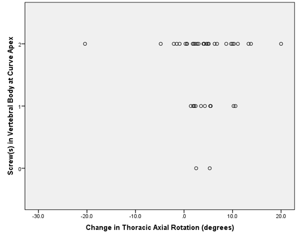
Table 4. Spearman correlation coefficient demonstrates no statistically significant relationship between percent change in axial rotation and screw location relative to the curve apex.
|
Correlations |
|
|
% Change |
|
Spearman's rho |
Percent Change |
Correlation Coefficient |
1.000 |
Sig. (2-tailed) |
- |
n |
41 |
|
Closest Screw Above Apex Thoracic |
Correlation Coefficient |
0.002 |
Sig. (2-tailed) |
0.989 |
n |
41 |
|
Number of Screws Above |
Correlation Coefficient |
-0.102 |
Sig. (2-tailed) |
0.524 |
n |
41 |
|
Closest Screw Below Apex Thoracic |
Correlation Coefficient |
-0.013 |
Sig. (2-tailed) |
0.936 |
n |
41 |
|
Total number of screws below |
Correlation Coefficient |
0.000 |
Sig. (2-tailed) |
1.000 |
n |
41 |
Adolescent idiopathic scoliosis is a condition that has historically been evaluated by frontal and lateral radiographs with the degree of coronal and sagittal curvature pre and postoperatively determined by Cobb angle. Recent advances in the understanding of scoliotic curves has highlighted the contribution of the third dimension, axial rotation, to the degree of thoracic distortion. The degree of axial rotation can be an indicator of curve progression and therefore measurements of axial rotation are of key significance in the prognosis and treatment of AIS [2,3,11-13].
This study utilized CT for preoperative and postoperative evaluation. CT scanning has been shown to be highly accurate for preoperative evaluation of axial rotation [3,4] and postoperative evaluation of screw breach and related surgical complications [14-16]. Radiographic techniques are most commonly employed for evaluation of vertebral rotation pre- and postoperatively due to their simplicity but are prone to interobserver variability and landmark obscuration by hardware [3].
It has been argued that CT scans are unable to correctly identify the curve apex in scoliotic patients due to their axial acquisition and display. This argument is no longer valid as the majority of CT scanners currently in use have the capability to create reformatted images. This study demonstrates the ease in identification of the apex vertebra on the coronal reformatted image with the ability to cross reference to the axial and sagittal planes (Figure 1). Helical CT scanning provides the ability to reformat in any plane, allowing more detailed evaluation of the anatomy preoperatively and hardware position postoperatively (Figure 2). Three-dimensional surface rendered images (Figure 3) and images with density thresholds optimized to display metallic hardware (Figure 4) can also be easily obtained for improved delineation of anatomy and hardware.
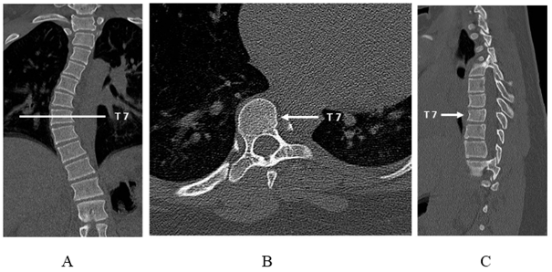
Figure 1: Pre-operative CT localization scan obtained in a 16-year-old girl with thoracic dextroscoliosis. The thoracic curve apex (T7) is localized on 2-dimension coronal reconstruction A and cross-referenced to the axial B and sagittal reconstruction C views at a dose equivalent to a 2-view spine radiograph series.
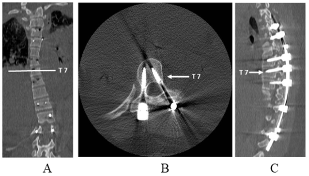
Figure 2. Post-operative CT scan obtained in the same patient allows detailed hardware evaluation in the coronal A, axial B and sagittal C planes.

Figure 3. 3D surface rendered images. Pre-operative A and post-operative B 3D volume surface rendered images can be rotated around the craniocaudal axis to best delineate the three-dimensional curvatures.
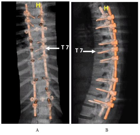
Figure 4. Post-operative 3D volume rendered images. Volumetric display of the postoperative spine with density thresholds optimized for hardware visualization (shown here in the frontal A and lateral B projections) can be rotated around the craniocaudal axis, improving evaluation of hardware positioning.
Multiple methods have been used to estimate the axial rotation by CT in patients with AIS. Our study utilized the Ho method [9] (Figure 5), which has close correlation to actual rotation values as well as clearly defined reference points with minimal interobserver variability [2,16]. Pedicle screw placement has become the preferred method to safely and effectively reduce rotation within coronal, sagittal and axial planes in patients with AIS [17-19]. This focus on effective reduction in axial rotation has coincided with the current preference for use of pedicle screw constructs for improved AIS correction. Hypoplastic and distorted pedicles may occur near in patients with AIS patients, particularly near the curve apex [7,8,17,20,21]. Pedicular variant anatomy and overall anatomic distortion by the scoliosis are concerning for potential neurovascular complication during surgery.
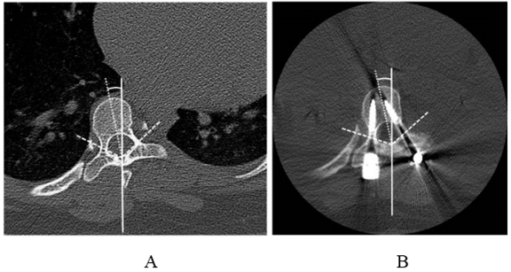
Figure 5. Ho method of measuring axial rotation angles on CT. Measurements are shown on pre-operative A and post-operative B images. Lines are drawn connecting a point at the laminar junction with the pedicle-lamina junction bilaterally (dashed lines), and the angle between these lines is bisected (dotted line). The rotation angle (curved line) is measured between the bisector and the vertical line. In this case, the rotation angle decreased by 0.6 degrees postoperatively.
Our study was aimed at determining whether instrumentation of these potentially unsafe levels was necessary for maximum correction of axial rotation. We have demonstrated that pedicle screw instrumentation at the apical thoracic vertebra does not correlate with improved correction in axial rotation. Additionally, surgical instrumentation of the three levels central to the thoracic curve apex (defined as the apical and two flanking vertebral bodies) does not correlate with improved axial rotation correction. This finding indicates that these potentially dangerous levels can be avoided without compromising the degree of correction in axial rotation.
We also evaluated the degree of axial rotation correction in relation to the total number of pedicle screws placed (screw density). We found no correlation between screw density and improvement in axial rotation postoperatively. While this finding seems counterintuitive, it has also been shown that screw density does not directly correlate with the degree of coronal and sagittal correction in cases of AIS [21,22]. Findings may implicate other inherent or anatomic factors such as curve flexibility [24] as more significant etiologies. Therefore, the use of high screw density with its increased costs and complication risk (of 15.7% per screw [25]) may be avoided.
Correction of axial rotation in AIS patients is important for trunk rotation and cosmetic deformity. The most deformed and hypoplastic pedicles in AIS are present at or near the curve apex where axial rotation is also maximal. Our study has shown that instrumentation of the pedicles at or near the thoracic apex level does not correlate with the degree of axial correction. The density of pedicle screws also had no correlation with decrease in axial rotation postoperatively. Therefore, instrumentation of the apex level and placement of an excessive number of screws could be avoided thereby decreasing both costs and complications related to screw breach.
The primary limitation of this study was that all surgical procedures were performed by the same surgeon. The findings of this study may not be representative of all spinal surgeons who may demonstrate significant variation with respect to skill and experience in spinal instrumentation for AIS.
The axial rotation of the spine of a patient with AIS may be reduced when the patient is supine [3]. Because computed tomography is acquired typically with the patient in the supine position, images may underestimate the severity of deformity among patients with AIS with respect to the axial plane. This variation, if present, may affect both pre- and postoperative CT scans and, therefore, not affect the overall change. Alternatively, the increased rigidity of the spine postoperatively may mean that the alteration mostly affects the preoperative scan, thereby underestimating the postoperative percent change in rotation.
Inexact patient positioning for CT scanning may affect the measurement of axial rotation obtained by the method outlined by Ho who describes difficulty in identifying reliable datum points for the calculation of rotation angle. Every method of axial rotation measurement has assumptions inherent to the method that cause some degree of inaccuracy [3]. This method was chosen as it has been shown to be equally accurate and to have less interobserver variability than other CT based methods [3].
We want to express our gratitude to Julie Pepe, PhD, for her contribution in the study’s statistical analysis.
L W Bancroft – Lippincott book royalties, Thieme Chief Editor travel assistance
- Atmaca H, Inanmaz ME, Bal E, Caliskan I, Kose KC (2014) Axial plane analysis of Lenke 1A adolescent idiopathic scoliosis as an aid to identify curve characteristics. Spine J. 14: 2425-2433. [Crossref]
- Lam GC, Hill DL, Le LH, Raso JV, Lou HE (2008) Vertebral rotation measurement: a summary and comparison of common radiographic and CT methods. Scoliosis. 3: 16. [Crossref]
- Kuklo TR, Potter BK, Lenke LG (2005) Vertebral rotation and thoracic torsion in adolescent idiopathic scoliosis: what is the best radiographic correlate? J Spinal Disord Tech. 18: 139-147. [Crossref]
- Ho EK, Upadhyay SS, Ferris L, Chan FL, Bacon-Shone J, et al. (1992) A comparative study of computed tomographic and plain radiographic methods to measure vertebral rotation in adolescent idiopathic scoliosis. Spine. 17: 771-774. [Crossref]
- Stokes IA, Bigalow LC, Moreland MS (1987) Three-dimensional spinal curvature in idiopathic scoliosis. J Orthop Res 5: 102-113. [Crossref]
- Xiong B, Sevastik J, Hedlund R, Sevastik B (1993) Segmental vertebral rotation in early scoliosis. Eur Spine J. 2: 37-41. [Crossref]
- Hu X, Siemionow KB, Lieberman IH (2014) Thoracic and lumbar vertebrae morphology in Lenke type 1 female adolescent idiopathic scoliosis patients. Int J Spine Surg. 1: 8. [Crossref]
- Sarwahi V, Sugarman EP, Wollowick AL, Amaral TD, Lo Y, et al. (2014) Prevalence, distribution, and surgical relevance of abnormal pedicles in spines with adolescent idiopathic scoliosis vs. no deformity: a CT-based study. J Bone Joint Surg Am. 96: e92. [Crossref]
- Ho EK, Upadhyay SS, Chan FL, Hsu LC, Leong JC (1993) New methods of measuring vertebral rotation from computed tomographic scans. An intraobserver and interobserver study on girls with scoliosis. Spine. 18: 1173-1177. [Crossref]
- Krismer M, Bauer R, Sterzinger W (1992) Scoliosis correction by Cotrel-Dubousset instrumentation. The effect of derotation and three dimensional corrections. Spine. 17: S263-S269. [Crossref]
- Drerup B (1985) Improvements in measuring vertebral rotation from the projections of the pedicles. J Biomech. 18: 369-378. [Crossref]
- Skalli W, Lavaste F, Descrimes JL (1995) Quantification of three-dimensional vertebral rotations in scoliosis: what are the true values? Spine. 20: 546-553. [Crossref]
- de Kleuver M, Lewis SJ, Germscheid NM, Kamper SJ, Alanay A, et al. (2014) Optimal surgical care for adolescent idiopathic scoliosis: an international consensus. Eur Spine J. 23: 2603-2618. [Crossref]
- Macke J, Woo R, Varich L (2016) Accuracy of robot-assisted pedicle screw placement for adolescent idiopathic scoliosis in the pediatric population. J Robot Surg. 10: 145-150. [Crossref]
- Farber GL, Place HM, Mazur RA, Jones DE, Damiano TR (1995) Accuracy of pedicle screw placement in lumbar fusions by plain radiographs and computed tomography. Spine. 20: 1494-1499. [Crossref]
- Göçen S, Aksu MG, Baktiroğlu L, Ozcan O (1998) Evaluation of computed tomographic methods to measure vertebral rotation in adolescent idiopathic scoliosis: an intraobserver and interobserver analysis. J Spinal Disord. 11: 210-214. [Crossref]
- Matsumoto M, Watanabe K, Hosogane N, Toyama Y (2014) Updates on surgical treatments for pediatric scoliosis. J Orthop Sci. 19: 6-14. [Crossref]
- Lee SM1, Suk SI, Chung ER (2004) Direct vertebral rotation: a new technique of three-dimensional deformity correction with segmental pedicle screw fixation in adolescent idiopathic scoliosis. Spine. 29: 343-349. [Crossref]
- Huang Z, Wang Q, Yang J, Yang J, Li F (2016) Vertebral derotation by vertebral column manipulator improves postoperative radiographs outcomes of Lenke 5C patients for follow up minimum 2 Year. Clin Spine Surg. 29: E157-161. [Crossref]
- Sugimoto Y, Tanaka M, Nakanishi K, Misawa H, Takigawa T, et al. (2007) Predicting intraoperative vertebral rotation in patients with scoliosis using posterior elements as anatomical landmarks. Spine. 32: E761-763. [Crossref]
- Watanabe K, Lenke LG, Matsumoto M, Harimaya K, Kim YJ, et al. (2010) A novel pedicle channel classification describing osseous anatomy: how many thoracic scoliotic pedicles have cancellous channels? Spine. 35: 1836-1842. [Crossref]
- Rushton PR, Elmalky M, Tikoo A, Basu S, Cole AA, et al. (2016) The effect of metal density in thoracic adolescent idiopathic scoliosis. Eur Spine J. 25: 3324-3330. [Crossref]
- Bharucha NJ, Lonner BS, Auerbach JD, Kean KE, Trobisch PD (2013) Low-density versus high-density thoracic pedicle screw constructs in adolescent idiopathic scoliosis: do more screws lead to a better outcome? Spine J. 13: 375-381. [Crossref]
- Chen J, Yang C, Ran B, Wang Y, Wang C, et al. (2013) Correction of Lenke 5 adolescent idiopathic scoliosis using pedicle screw instrumentation: does implant density influence the correction? Spine. 38: E946-951. [Crossref]
- Hicks JM, Singla A, Shen FH, Arlet V (2010) Complications of pedicle screw fixation in scoliosis surgery: a systematic review. Spine. 35: E465-470. [Crossref]


